SonoStudy: Ultrasound of heart and lungs identify preeclamptic pregnant patients at risk

In anesthesiology literature (not yet on pubmed), heart and lung ultrasound has entered the mix to identify pregnant patients at risk for respiratory failure – looking for hypocontractile heart and B lines! Oh yeah! It has received a lot of press, see here, here and here :

“Chicago — (March 18, 2014)

An ultrasound of the lungs could help doctors quickly determine if a pregnant woman with preeclampsia is at risk for respiratory failure, suggests preliminary research published in the April issue of Anesthesiology.

About 60,000 women worldwide die as a result of preeclampsia, which causes severely high blood pressure. Potential complications include stroke, bleeding and excess fluid in the lungs – called pulmonary edema – which can lead to respiratory failure.  The study suggests a lung ultrasound can help doctors easily learn whether a woman with preeclampsia is suffering from pulmonary edema and ensure she receives the correct treatment.

“Lung Ultrasound is fast, safe, noninvasive and easy to use,” said Marc Leone, M.D., Ph.D., lead author of the study and vice chair of the department of anesthesiology and critical care medicine, Hopital Nord, Marseille, France. “We found it allowed us to quickly assess whether a woman with preeclampsia had pulmonary edema and confirm the severity of the condition.”

Doctors often measure urine output to determine if a woman needs fluid administration, but the results are wrong about half of the time. “Lung ultrasound enables the medical team to identify which women really need the fluid treatment,” noted Dr. Zieleskiewicz, the study’s  first author.

Pulmonary edema is typically caused by heart failure, but also can be caused by lung inflammation. Researchers analyzed the use of lung ultrasounds, which can assess lung  edema, are easier to use than cardiac ultrasound and can be performed with devices commonly found in maternity wards. Lung ultrasound highlights white lines mimicking comet tails, irradiating from the border of the lungs. These lines are the reflection of water in the lungs.  The detection of three or more lines strongly suggests the diagnosis of pulmonary edema.

Researchers performed both cardiac and lung ultrasounds before and after delivery in 20 women with severe preeclampsia.  Five of the 20 women (25 percent) had pulmonary edema prior to delivery according to lung ultrasound, while four (20 percent) had the condition according to the cardiac ultrasound. The lung ultrasound identified a patient with non-cardiac pulmonary edema, which the cardiac ultrasound did not detect.

The test results could help ensure that pregnant women with pulmonary edema not be given intravenous or excess fluids, which worsens the condition and can lead to respiratory failure.  Typically, women with pulmonary edema are treated with oxygen and medication to lower the blood pressure or rid the body of excess fluid.  In real time, lung ultrasound also serves to observe improvement or worsening of pulmonary edema.”

SonoCase: Thoracic aortic aneurysms – and a review of the literature #FOAMed

Thoracic aortic aneurysms are much less common than abdominal aortic aneurysms. And, to top it off, the measurements of the thoracic aorta are different than the abdominal aorta, especially in the ascending thoracic aorta. It’s good to know how to look at the thoracic aorta when you need to, and what the normal measurements are so that you can make that immediate decision when a thoracic aneurysm (or dissection) is detected.

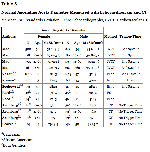
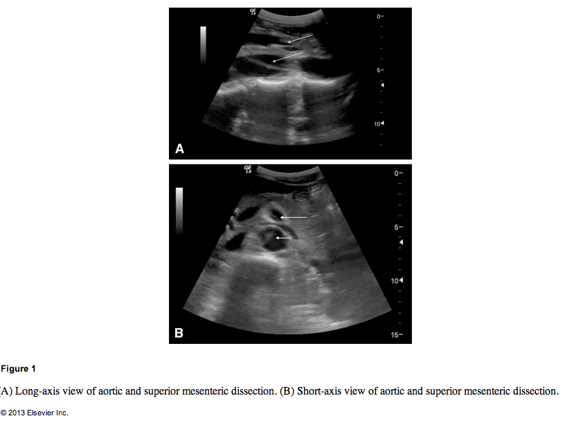
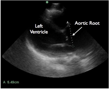
Drs. Daignault, Saul, and Lewiss published 2 great case reports in the Journal of EM Aug 2013 issue (subscription needed) on thoracic aortic aneurysms. One patient was a 60 year old male who had blunt trauma to his back by a heavy piece of plaster while at work. He had a BP of 140/80 mm Hg and HR of 90 beats/min. The FAST was negative for free fluid but the aortic root was found to be 5.49cm. This caused them to order a CT angio confirming their findings.  Another patient was an 82 year old male with 1 month of chest pain radiating to his back with history of aortic valve repair and HTN on coumadin. BP was 210/90 mm Hg. A bedside echo was performed and showed descending thoracic aorta aneurysm measuring 4.82cm.  This had a CT Angio done confirming descending thoracic aneurysm.  The abstract is found here, and a prior post on these case reports can be found here.

These cases bring up an excellent reason to review the thoracic aorta. The best cardiac echo view to see visualize the thoracic aorta, if you could only pick one, would be the parasternal long view.

According to a german study where they looked at the thoracic aorta throughout life from 17 to 89 yrs of age  via helical CT with the following methods and results: “Methods: Seventy adults, 17 to 89 years old, without any signs of cardiovascular disease were investigated with helical computed tomography. Aortic diameters were measured at seven predefined thoracic levels. Results: Aortic diameters (mean ± SD) were 2.98 ± 0.46 cm at the aortic valve sinus, 3.09 ± 0.41 cm at the ascending aorta, 2.94 ± 0.42 cm proximal to the innominate artery, 2.77 ± 0.37 cm at the proximal transverse arch, 2.61 ± 0.41 cm at the distal transverse arch, 2.47 ± 0.40 cm at the isthmus, and 2.43 ± 0.35 cm at the diaphragm. Men had slightly longer diameters than did women. All diameters increased with age. There was no influence of weight, height, or body surface area.”

More recently, Medscape authors state that the thoracic aorta dimensions are larger than the abdominal aorta dimensions and that aneursymal definition occurs when it is greater than 50% of the normal size.

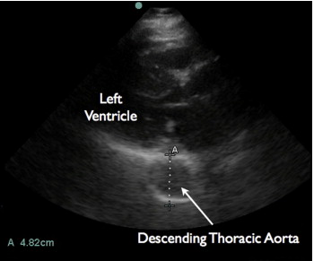
Another study in the radiology literature with many more subjects more recently, had the following methods and results: “Methods: 1442 consecutive subjects who were referred for evaluation of possible coronary artery disease underwent coronary CT angiography (CTA) and coronary artery calcium scanning (CACS) (55+11 years, 65% male) without known coronary heart disease, hypertension, chronic pulmonary and renal disease, diabetes and severe aortic calcification. The ascending aortic diameter, descending aortic diameter (DAOD), pulmonary artery (PAD) and chest anterioposterior diameter (CAPD), posterior border of sternal bone to anterior border of spine, were measured at the slice level of mid right pulmonary artery by using end systolic trigger image. The volume of four chambers, ejection fraction of left ventricle, and cardiac output were measured in 56% of the patients. Patients demographic information, age, gender, weight, height and body surface area (BSA), were recorded. The mean value and age specific and gender adjusted upper normal limits (mean + 2 standard deviations) were calculated. The linear correlation analysis was done between AAOD and all parameters. The reproducibility, wall thickness and difference between end systole and diastole were calculated. Result: AAOD has significant linear association with age, gender, descending aortic diameter and pulmonary artery diameter (P<0.05). There is no significant correlation between AAOD and body surface area, four chamber volume, LVEF, CO and CAPD. The mean Intra-luminal AAOD was 31.1 ± 3.9mm and 33.6 ± 4.1 mm in females and males respectively. The upper normal limits (mean + 2 standard deviations) of Intra-luminal AAOD, mean+ standard deviation, was 35.6, 38.3 and 40 mm for females and 37.8, 40.5 and 42.6 mm for males in age group 20 to 40, 41 to 60, above 60 year respectively. Intra-luminal should parallel echocardiography and invasive angiography. Traditional cross sectional imaging (with computed tomography and magnetic resonance imaging) includes the vessel wall. The mean total AAOD was 33.5mm and 36.0 mm in females and males respectively. The upper normal limits (mean + 2 standard deviations) of Intra-luminal AAOD, mean+ standard deviation, was 38.0, 40.7 and 42.4 mm for females and 40.2, 42.9 and 45.0 mm for males in age group 20 to 40, 41 to 60, above 60 year respectively. The inter and intra observer, scanner and repeated measurement variability was low (R value >0.91, P<0.001, coefficient variation <3.2%). AAOD was 1.7 mm less in end-diastole than end systole(P<0.001).” Below is their table illustrating the various measurements by others:

Screen Shot 2013-09-03 at 10.59.27 AM

As Dr Lewiss and colleagues state in their case reports, “The most recent consensus statement by the American Society of Echocardiography (ASE) and the American College of Emergency Physicians (ACEP) recognized that the thoracic aortic pathology can be identified on EP-performed focused cardiac ultrasound (3). Transthoracic echocardiography was shown to be consistent with TEE for measurement of the ascending aorta (21). Taylor et al. demonstrated that EP-performed focused cardiac ultrasound was consistent with CTA measurements for maximal thoracic aortic diameter (2). In these cases, an EP with considerable experience in ultrasound performed these studies, but many EP are trained in the use of focused ultrasound for evaluation of the abdominal aorta. Principals used in this application, such as avoiding measurements in oblique planes, measuring from outer wall to outer wall (for the descending thoracic aorta), and the use of Doppler also may be utilized in the evaluation of the thoracic portion of the aorta.”

SonoCase: 46yo c/o abdominal pain h/o cocaine use – sup mesenteric art dissection #FOAMed

Drs. Davis and Kendall write up a very interesting case in the Aug 2013 issue of Journal of EM where the ultrasound made the diagnosis, quite easily too. They discuss a 46 year old male with a history of current cocaine use AND a prior history of an aortic dissection, of course, who was complaining of sudden onset of abdominal pain and found to be severely hypertensive. The diagnosis on the top of their list was aortic dissection/aneurysm/rupture – and when they looked, they saw even more. Below is the abstract:

Background

A timely diagnosis of aortic dissection is associated with lower mortality. The use of emergent bedside ultrasound has been described to diagnose aortic dissection. However, there is limited literature regarding the use of bedside ultrasound to identify superior mesenteric artery dissection, a known high-risk feature of aortic dissection.

Objective

Our aim was to present a case of superior mesenteric artery dissection identified by bedside ultrasound and review the utility of bedside ultrasound in the diagnosis of aortic emergencies.

Case Report

We report a case of superior mesenteric artery dissection found on emergent bedside ultrasound in a 46-year-old male complaining of abdominal pain with a history of cocaine abuse and prior aortic dissection. Bedside ultrasound in the emergency department revealed an intimal flap in the descending aorta with extension into the superior mesenteric artery prompting early surgical consultation before computed tomography because of concern for acute mesenteric ischemia.

Conclusion

Superior mesenteric artery dissection is a high-risk feature of aortic dissection and can be identified with emergent bedside ultrasound.

Just one of their images is displayed below – but take a look at the video in JEM to truly see the awesomeness. A subscription and password is required, but it’s a great journal with lots of cool ultrasound cases published almost every month.

Screen Shot 2013-09-03 at 9.54.59 AM

SonoCase: 60yo blunt chest trauma, 82yo with chest pain- in JEM by @ultrasoundREL

In a recent article in the Journal of Emergency Medicine, Dr. Resa Lewiss and friends, discuss 2 cases of thoracic aortic aneurysm identification by focused cardiac ultrasound. It is a great case report that highlights the need to include the aortic root and descending thoracic aorta in the parasternal long view of your focused cardiac echo.

“A 60-year-old man presented to the emergency department (ED) after a blunt traumatic injury to his back while at work. During the focused cardiac ultrasound examination, the aortic outflow tract distal to the aortic valve appeared enlarged and the aortic root measured 5.49 cm.

Screen Shot 2014-03-04 at 9.16.55 AM

“An 82-year-old man with hypertension presented to the ED with 1 month of chest pain radiating to the back. The focused cardiac ultrasound examination demonstrated enlargement of the descending thoracic aorta at 4.82 cm.”

Screen Shot 2014-03-04 at 9.17.06 AM