Unknown's avatar

About SonoSpot

US Director Emeritus, Stanford Emergency Medicine Process Improvement Director, Stanford Emergency Medicine Co-Chair, Case Review Committee, Stanford Emergency Medicine

SonoStudy: Contrast Enhanced Ultrasound – the future for trauma assessment? #FOAMed

In a recent article in Critical Ultrasound Journal from July 2013, the authors (Italians, of course! – they always do things ahead of everyone!) discuss the utility of contrast enhancement for solid organ evaluation in trauma patients. So, the FAST scan will assess for free fluid from injury, but we dont know what that injury is through a simple FASt scan. With contrast, we can better visualize the solid organs and assess for injury. The authors say it best, “Computed Tomography (CT) is the standard reference in the emergency for evaluating the patients with abdominal trauma. Ultrasonography (US) has a high sensitivity in detecting free fluid in the peritoneum, but it does not show as much sensitivity for traumatic parenchymal lesions. The use of Contrast-Enhanced Ultrasound (CEUS) improves the accuracy of the method in the diagnosis and assessment of the extent of parenchymal lesions. Although the CEUS is not feasible as a method of first level in the diagnosis and management of the polytrauma patient, it can be used in the follow-up of traumatic injuries of abdominal parenchymal organs (liver, spleen and kidneys), especially in young people or children.”

The thing to keep in mind is that this is actually not new – but evolving and getting spoken about more and more – as the authors state: “The first results in the literature indicates the use of CEUS in patients with blunt abdominal trauma after the FAST (Focused Assessment with Sonography in Trauma) or the US, in hemodynamically stable patients with a history of low-energy trauma [1,4,6]. CT is reserved in cases of severe trauma, with clinical suspicion of multiorgan lesions and cases with inconclusive CEUS [6].”

How does contrast work sonographically? Read on : “The contrast agents eco-amplifiers are able to modify the acoustic impedance of tissues, interacting with ultrasound beams and increasing the echogenicity of the blood. The contrast media (CM) ultrasound (USCA, UltraSound Contrast Agent) consist of microbubbles containing inert gases and surrounded by membrane stabilizers. The power of echogenic microbubbles and acoustic impedance depends on the size of the microbubbles. The microbubbles, unlike the tissues and the free gas, are not simply passive reflectors, but expand and compress in response to the stages of compression and rarefaction of the acoustic wave, with increasingly large hikes in diameter. The non-linear oscillation of microbubbles determines the emission of frequencies of said second harmonic with a frequency which is twice the insonation. Through the use of specific software, low acoustic pressures and an algorithm of specific processing, it is possible to selectively display the signals from the CM, separating the signal of the microbubbles from the one regarding the tissue. This particular signal is identified in real time by means of two main algorithms: Pulse Inversion (PI) and Contrast Pulse Sequence (CPS) [7,8]”

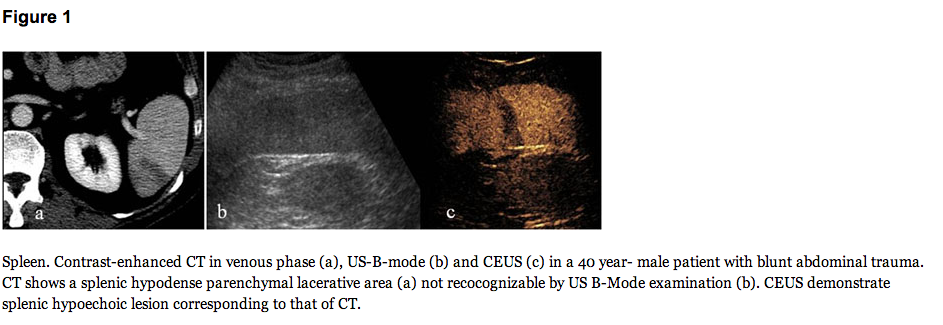
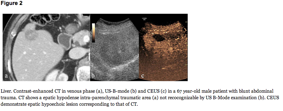
Here are some images from the authors in the article that makes the point:

Screen Shot 2013-08-27 at 7.27.41 PM Screen Shot 2013-08-27 at 7.28.21 PM

The conclusion? What to make of all of this?: “In the low-energy trauma and in hemodynamically stable patients, the US can be used as a first-level examination; when US detect intra-abdominal fluid CT examination is need. In the high-energy trauma the use of US as first line diagnostic is superfluous and damaging and the use of CT without and with i.v.c onstrast material is imperative. In order to reduce the radiation dose, particularly in young people or children, CEUS has an important role in the follow-up of conservatively treated traumatic injuries of the abdominal parenchymatous organs (liver, spleen and kidneys) diagnosed by CT [39,40]”

Read the article to get even more details on how the future of ultrasound will be, hopefully…here.

SonoEquipment: SonoSite’s new XPorte and their free teaching files on iTunes #FOAMed

SonoSite has launched a new ultrasound system called XPorte – it is an ultrasound machine and more! It’s all touch screen and not only allows for quick scanning and easy inputting of data, but it has lectures that you can view and listen to right there!

Screen Shot 2013-11-25 at 7.18.56 PM

You can download the free lectures and Xporte info from iTunes here. It’s another great resource for free ultrasound education. Hopefully SonoSite will not change that, but I doubt they will, as they already have a great app for free, called SonoAccess, as well as awesome online lecture tutorials by the best in the field in their online learning center.

SonoGlobalHealth: What Ultrasound does in Uganda to prevent MTCT of HIV #FOAMed #FOAMus

A friend of mine, William Cherniak – a family medicine resident in Canada at the University of Toronto-  has been working on a project. Not just any project, but a global impact project with the help of KIHEFO (Kigezi Healthcare Foundation) and his own group that he created called TO-the World. What is this project, you ask? Well, it was to show how minimizing maternal to child transmission (MTCT) of HIV can be helped by simply having and letting the women know in the region that a portable ultrasound has arrived. This project will be presented at Global Health 2013

Screen Shot 2013-08-29 at 5.50.19 PM

The project in brief: “In
 alignment
 with 
the 
WHO 2010 
guidelines 
for
 MTCT elimination and 
the
 Ugandan 
government’s
 adoption
 of plan
B+; KIHEFO,
 in
 collaboration 
with
 TO
– the
WORLD,
 designed
 a structured maternal
 health
camp [sMHC]
 centered on providing
 expectant
 mothers in
 rural
 Uganda with a 
free
 obstetric
ultrasound [OBU]. The
 four‐pronged
 approach
 of
 MTCT
 elimination 
was
 followed 
in
 the
 design
 of 
the 
sMHC. In 
one 
day, 
45 
women rotated 
through
 registration, 
pre‐test
 counseling, 
testing
 for 
HIV 
and 
Syphilis, 
family
 planning,
 obstetric
 ultrasound 
and, 
for
the 
women 
identified 
as
 being 
high 
risk
 by 
triage, 
dental 
and/or 
medical 
services. 
Each
 woman
 received 
fansidar, 
multivitamins,
 folic
acid
 and
 filled 
in 
a 
pre‐ designed
questionnaire.
 In 
total,
10 women
 identified 
themselves 
as 
being 
HIV+
 at 
registration, surprisingly 
only 
half 
were currently being 
treated 
with 
ARVs. 

An 
additional 
two 
women
 were 
diagnosed 
as 
being 
HIV+ 
during 
the 
health
camp.

 All
 women 
received 
counseling
 and
 were
 started
 on
 ARVs for 
life. Only 
7 
women 
had 
ever 
previously 
had 
an 
OBU, 
and
all
 45 
women 
verbally 
identified 
that 
the 
reason 
for
 attending 
the 
antenatal 
health
camp 
was 
to 
receive 
a 
free 
OBU.” – Now that is just amazing. The power of what a portable ultrasound can have on a community in fighting MTCT of HIV. Bring the ultrasound, and they will come – that is your way to start the healthcare and assessment they need.

They continue to state how they hope to solve the problem of high maternal death and high infant death rates: ” Solving
 the 
Problem
– Ultrasound
 and 
Outreach 
to 
Reduce
 Maternal 
Mortality. As
 stated 
above, 
the 
leading 
causes
 of 
maternal
 mortality 
include  hemorrhage, 
eclampsia, 
obstructed 
labor,
 infections,
 and
 birth 
defects. 

Studies 
have 
shown 
that 
obstetric 
ultrasound
 imaging 
can 
prevent 
most outcomes
 by 
providing 
early 
diagnosis 
and 
intervention. 

By 
providing useful 
information 
such 
as 
whether 
or 
not 
the 
mother 
is 
carrying 
twins, 
has 
an 
ectopic 
pregnancy 
or 
otherwise a 
mother 
and
her
 partner 
can
 make
 an 
informed 
decision 
about 
whether 
or 
not 
to 
deliver 
at 
home 
with 
untrained
 professionals, or 
a
 health
center where 
they 
can receive 
life‐saving
 treatment. Furthermore, 
the 
World 
Health 
Organization
 (2003)
 recognizes 
ultrasound 
technology 
as 
ideally 
suited
 to 
low
 and 
middle 
income countries, 
as 
it 
is
 relatively 
low‐cost, 
low
 input, 
and
 easily 
maintained
 and 
transported. 

Additionally, 
studies
 conducted
 on
 the 
use 
of 
ultrasound 
technology 
in 
two 
rural 
hospitals 
in 
Rwanda
 have 
indicated 
that 
after 
an 
initial
 training 
period,
 an 
ultrasound
 program 
led 
by 
local 
health 
care 
providers 
can 
be 
sustainable
 and 
lead 
to
 accurate 
diagnoses. Ultrasound 
imaging 
is 
beneficial 
to 
rural 
populations 
as 
it 
is 
a
 simple 
a
nd non‐invasive
 procedure. This
 helps 
to 
reduce levels 
of 
fear 
from 
women 
who 
have previously maintained 
their 
cultural
 preferences 
for 
receiving 
treatment 
and
 giving 
birth 
with 
untrained 
birth 
attendants 
in 
their 
local
 villages.

Sustainability: Various
 studies 
on
 obstetric 
ultrasound 
imaging 
as 
a 
sustainable 
and 
appropriate 
technology 
to 
developing
 nations, 
and 
its 
capacity 
to 
reduce 
rates 
of 
maternal 
mortality, 
have 
been 
conducted 
in 
rural
 regions
 of 
Rwanda, 
Botswana
 and 
Tanzania. 
In 
northern 
Tanzania, 
a 
study 
conducted 
amongst
 women
 who
 were
receiving 
ultrasound 
imaging
 for 
the 
first 
time 
indicated 
that 
the 
majority 
of 
women 
were 
satisfied 
with the 
information 
provided 
from 
the 
procedure, 
despite 
not 
initially  understanding 
its 
purpose. 
The 
ultrasound
 procedure
 provided 
women 
with 
the 
ability 
to 
see 
fetal 
positioning,
 fetal 
sex, 
and
 to 
recognize 
any 
potential
 pregnancy
complications. 
Information,
 particularly 
the 
latter, 
resulted 
in 
guiding
 treatment
 for 
the
woman’s
 particular birth
– helping 
her
 make
 an
 informed
 decision
 as 
to 
where
 and
 how 
she 
would
 deliver.
 This
 particular 
study
 in
 Tanzania 
concluded
 that
 the 
ultrasound 
imaging 
was 
useful
 in 
reducing 
the 
risk 
of
 maternal
 mortality, 
although
 the 
treatment 
should 
be 
accompanied 
by 
a 
thorough
 education 
campaign 
and 
consent program.”

The future? “Currently,
 TO
– the
WORLD 
is 
in 
the 
process 
of 
raising 
funds 
to 
purchase 
two 
portable 
ultrasound 
machines.  

These 
machines 
will 
be 
purchased 
locally 
in 
Uganda 
to 
ensure 
sustainability 
in 
maintenance 
and 
economic 
stimulus for 
the 
communities 
in 
which 
they 
serve. Multiple 
outreach 
camps
 will 
be 
conducted 
in 
2014 
with 
the 
previously 
designed 
model 
based 
on 
the 
WHO 
four‐pronged 
approach 
to 
MTCT 
elimination 
of 
HIV.

The WSJ actually spoke of what happens when an US machine is taken to a developing country – more antenatal visits!

Here is the video William made to support his cause:

1. Report
of
a
WHO
Technical
Consultation.
Towards
the
elimination
of
mother‐to‐child
transmission
of
HIV.

Accessed
March,
2013
at
http://www.who.int/hiv/pub/mtct/elimination_report/en/index.html
2. IRIN
Humanitarian
News
and
Analysis
–A
service
of
the
UN
office
for
the
coordination
of
human
affairs.

Accessed
March,
2013
at
http://www.irinnews.org/Report/96308/UGANDA‐Government‐
adopts‐new‐PMTCT‐strategy
3. WHO
Executive
Summary,
April 2012.
Use
of
Antiretroviral
Drugs
for
Treating
Pregnant
Women
and
Preventing
HIV
Infections
in
Infants.

Accessed
March,
2013
at

Click to access PMTCT_update.pdf

4. World
Health
Organization. Statistics
on
Maternal
Mortality
in
Uganda,
accessed
March,
2013
at
http://www.who.int/healthinfo/statistics/indmaternalmortality/en/index.html
5. Kigezi
Healthcare
Foundation
website,
accessed
March,
2012
at
www.kihefo.org
6. Maternal
Health:
Investing
in
the
Lifeline
of
Healthy
Societies
and
Economies.
Africa
Progress
Panel
Position
Piece.
September
2010.
7. Yaw
A.W.,
Alexander
T.O.,
and
Edward
T.D.
The
Role
of
Obstetric
Ultrasound
in
Reducing
Maternal
and
Perinatal
Mortality,
Ultrasound
Imaging
‐ Medical
Applications,
InTech,
Accessed
March,
2013.
Available
from:
http://www.intechopen.com/books/ultrasound‐imagingmedical‐applications/the‐
role‐of‐obstetric‐ultrasound‐in‐reducing‐maternal‐and‐perinatal‐mortality.
8. Shah
S.P.,
Epino
H.,
Bukhman
G.,
Umulisa
I.,
Dushimiyimana
J.M.,
Reichman
A.,
Noble
V.E.
Impact
of
the
introduction
of
ultrasound
services
in
a
limited resource
setting:
rural
Rwanda.
BMC
InternationalHealth
Human
Rights.
2009;27:9‐4
9. Firth
E.R.,
Mlay
P.,Walker
R.,
Sill
P.R.
Pregnant
women’s
beliefs,
expectations
and
experiences
of
antenatal
ultrasound
in
Northern
Tanzania.African
Journal
of
Reproductive
Health.
2011;
15(2):91‐
107

SonoStudy: Ultrasound differentiating perforated from non-perforated appendicitis #FOAMed #FOAMus

In a study published in AJR, a very hot topic was reviewed. 2 centers. 160 kids. Ultrasound and appendectomy with comparison to operative report. Do I have your attention now? This is a tough one, ultrasound for appendicitis is being recommended by pediatricians, radiologists, emergency physicians and surgeons. A big limitation was thought that ultrasound is not great for differentiating perforated from non-perforated appendicitis…. in addition to other limitations including bowel gas scatter limiting view of the entire appendix, and variations in appendix size that may have a false positive for appendicitis if diameter size alone is used as the indicator. Well, it isnt perfect – we know that.

Now, to review, appendicitis is diagnosed by applying the linear (or curvilinear if added depth is needed) probe to the area where the patient points to noting maximal pain, with the indicator toward the patient’s right side. Graded compression is then performed in that region which should displace and flatten bowel, identifying the psoas muscle and the transverse view of the iliac vessels. The appendix usually is located just anterior to these structures coming off of the cecum, and is normally compressible without being more than 6mm in diameter. It may be in its transverse or longitudinal view depending on anatomy. The entire appendix should be viewed, including to its tip. Be sure to view it in two orthogonal planes (rotate probe 90 degrees) to ensure it is the appendix, as a lymph node may look very similar to a transverse appendix but will not elongate into a tubular structure when viewed in its longitudinal plane. Here are some views of a positive appendicitis (absence of compressibility with attempts, dilated appendix):

APPENDICITIS WITH MEASUREMENTS_crop

Appendicitis by Ultrasound: A greater than 6mm in diameter, aperistaltic, non-compressible appendix +/- appendecolith.

Ultrasound Podcast posted a great video a year ago on the “how-to” for appendix ultrasound and why to go to ultrasound first in the work up of appendicitis:

Let’s go back to the study:

“OBJECTIVE. Acute appendicitis is the most common condition requiring emergency surgery in children. Differentiation of perforated from nonperforated appendicitis is important because perforated appendicitis may initially be managed conservatively whereas nonperforated appendicitis requires immediate surgical intervention. CT has been proved effective in identifying appendiceal perforation. The purpose of this study was to determine whether perforated and nonperforated appendicitis in children can be similarly differentiated with ultrasound.

MATERIALS AND METHODS. This retrospective study included 161 consecutively registered children from two centers who had acute appendicitis and had undergone ultra-sound and appendectomy. Ultrasound images were reviewed for appendiceal size, appearance of the appendiceal wall, changes in periappendiceal fat, and presence of free fluid, abscess, or appendicolith. The surgical report served as the reference standard for determining whether perforation was present. The specificity and sensitivity of each ultrasound finding were determined, and binary models were generated.

RESULTS. The patients included were 94 boys and 67 girls (age range, 1-20 years; mean, 11 ± 4.4 [SD] years) The appendiceal perforation rate was significantly higher in children younger than 8 years (62.5%) compared with older children (29.5%). Sonographic findings associated with perforation included abscess (sensitivity, 36.2%; specificity, 99%), loss of the echogenic submucosal layer of the appendix in a child younger than 8 years (sensitivity, 100%; specificity, 72.7%), and presence of an appendicolith in a child younger than 8 years (sensitivity, 68.4%; specificity, 91.7%).

CONCLUSION. Ultrasound is effective for differentiation of perforated from nonperforated appendicitis in children.”

Interestingly, a multi-organizational group came together for guidelines published in a study in Pediatric Emergency Care. : abstract below:

“The objective of this study was to compare usage of computed tomography (CT) scan for evaluation of appendicitis in a children’s hospital emergency department before and after implementation of a clinical practice guideline focused on early surgical consultation before obtaining advanced imaging.

METHODS:

A multidisciplinary team met to create a pathway to formalize the evaluation of pediatric patients with abdominal pain. Computed tomography scan utilization rates were studied before and after pathway implementation.

RESULTS:

Among patients who had appendectomy in the year before implementation (n = 70), 90% had CT scans, 6.9% had ultrasound, and 5.7% had no imaging. The negative appendectomy rate before implementation was 5.7%. In patients undergoing appendectomy in the postimplementation cohort (n = 96), 48% underwent CT, 39.6% underwent ultrasound, and 15.6% had no imaging. The negative appendectomy rate was 5.2%. We demonstrated a 41% decrease in CT use for patients undergoing appendectomy at our institution without an increase in the negative appendectomy rate or missed appendectomy. The results were even more striking when comparing the rate of CT scan use in the subset of patients undergoing appendectomy without imaging from an outside hospital. In these patients, CT scan utilization decreased from 82% to 20%, a 76% reduction in CT use in our facility after protocol implementation.

CONCLUSIONS:

Implementation of a clinical evaluation pathway emphasizing examination, early surgeon involvement, and utilization of ultrasound as the initial imaging modality for evaluation of abdominal pain concerning for appendicitis resulted in a marked decrease in the reliance on CT scanning without loss of diagnostic accuracy.”

Why talk about this? Well, there is ALWAYS, always, ALWAYS press about how ultrasound can and should be used for appendicitis evaluation in pediatric patient for radiation exposure minimization. It does have false negatives and false positives though – as with all thing ultrasound, you must know it’s strengths and weaknesses….and correlate clinically 🙂

SonoStudy: Ultrasound for shoulder dislocation – Dx to anesthesia & reduction #FOAMed #FOAMus

A recent study in Annals of Emergency Medicine (found on pubmed too) discusses the use of ultrasound for assessing shoulder dislocation and reduction. Yup, that’s right – no need for that Xray – unless you are concerned about a fracture. But, when you have a patient with a history of shoulder dislocation saying, “it’s out again” then dont get that Xray – before or after your reduction – just use ultrasound. It’s quick and easy and can also be used for joint injections for anestheisa too. Dr. Mike Stone showed a great video of this too – 2 docs competing to see who finishes the assessment, anesthesia and reduction the quickest – guess who won….

Diagnostic Accuracy of Ultrasonographic Examination in the Management of Shoulder Dislocation in the Emergency Department

Study objective

Emergency physicians frequently encounter shoulder dislocation in their practice. The objective of this study is to assess the diagnostic accuracy of ultrasonography in detecting shoulder dislocation and confirming proper reduction in patients presenting to the emergency department (ED) with possible shoulder dislocation. We hypothesize that ultrasonography could be a reliable alternative for pre- and postradiographic evaluation of shoulder dislocation.

Methods

This was a prospective observational study. A convenience sample of patients suspected of having shoulder dislocation was enrolled in the study. Ultrasonography was performed before and after reduction procedure with a 7.5- to 10-MHz linear transducer. Shoulder dislocation was confirmed by taking radiographs in 3 routine views as a criterion standard. The operating characteristics of ultrasonography to detect dislocation in patients with possible shoulder dislocation and to confirm reduction in patients with definitive dislocation were calculated as the primary endpoints.

Results

Seventy-three patients were enrolled. The ultrasonography did not miss any dislocation. The results of ultrasonography and radiography were identical and the sensitivity of ultrasonography in detection of shoulder dislocation was 100% (95% confidence interval 93.4% to 100%). The sensitivity of ultrasonography for assessment of complete reduction of the shoulder joint reached 100% (95% confidence interval 93.2% to 100%) in our study as well.

Conclusion

We suggest that ultrasonography be performed in all patients who present to the ED with a clinical impression of shoulder dislocation on admission time. The results of this study provide promising preliminary support for the ability of ultrasonography to detect shoulder dislocation. However, further investigation is necessary to validate the results and assess the ability of ultrasonography in detecting fractures associated with dislocation.

To view Dr. Mike Stone’s lecture on shoulder dislocation diagnosed by ultrasound, view below:

For another great post of shoulder shrugging – see broomedocs site here!

ACEP News in 2/2014 had an article on shoulder dislocation by ultrasound – go here.

SonoCase: 25yo unresponsive, found down – by @KasiaHamptonMD #FOAMed #FOAMus

In case you all were unaware, Dr. Kasia Hampton is REALLY into ultrasound. She is a resident in emergency medicine and is teaching her colleagues how to use it. She has case after case of great findings, quick pick-ups, and lives saved and management changed due to that little old ultrasound machine. She even has another twitter/blog, called @tres_EUS  – a site for residents interested in ultrasound cases/leadership/research/etc. She emailed me this case that I thought was a fabulous use of ultrasound and actually shows what I harped on and on about with EMCrit on a recent podcast on FAST scans highlighted in our SonoTips and Tricks on FAST scan upper quadrants.

Enjoy!

“25 yo male was found unresponsive per bystanders. Upon EMS arrival he was noted to have multiple stab wounds to the upper extremities and chest. Initial set of vitals revealed tachycardia without hypotension. Patient was intubated at the scene “for airway protection”. Mechanically ventilated upon ED arrival with the following vitals: BP 135/90 mmHg, HR 105 BPM, respirations 16/min, SpO2 100%, T 35.8 C. GCS 3T. During secondary survey found to have one stab wound to the left anterior chest (inferior to the nipple), and second stab wound to the right posterior chest (lateral to the inferior aspect of the scapula). Additional two stab wounds to both shoulders were superficial and were no longer bleeding. No apparent abdominal (wall) injuries were noted. Abdomen was non-distended and soft.

The RUQ FAST scan:

Seek and ye shall find 3

FAST ultrasound evaluation was performed after the patient was log-rolled in both directions – first to the left and then to the right.  Subsequently the patient was taken to CT scan. He remained hemodynamically stable. Below the comparative findings of FAST vs CT scans.

IMAGING

FAST ULTRASOUND

CT

RUQ

perihepatic free fluid

perihepatic free fluid

SUBXIPHOID

no pericardial effusion

no pericardial effusion

LUQ

no free fluid

trace perisplenic free fluid

PELVIC

no free fluid

no free fluid

Given stab wound to left anterior chest with presence of free fluid in the abdomen (with hepatic and splenic injuries identified on CT), patient was taken to the operating room. Injury to pericardium itself without pericardial effusion was suspected on CT. During the surgical exploration it appeared that the stab wound to the left chest only nicked the pericardium (no blood within pericardial sac), while penetrating the left diaphragm, left lobe of the liver, stomach, spleen and pancreatic body.

This case illustrates a few important concepts:

  1. The ultimate importance of visualizing the paracolic gutter around inferior pole of the right kidney on FAST ultrasound exam;
  2. The dilemma of performing FAST scans after the patient has been log-rolled (in particular to the left side, while less important if rolled onto the right);
  3. The superiority of Secondary UltraSonographic Survey In Trauma (SUSS IT) over clinical exam for non-suspected injuries.

4 @broomedocs with love - SUSS IT OUT

In this particular case I wonder if the trace perisplenic free fluid would have been identified on FAST performed before log-rolling? Additionally, it is quite amazing how misleading was the clinical secondary survey in comparison to FAST findings and intra-operative discoveries. “

SonoCase: 72yo back pain & hypotensive – by Dr. Calvin Hwang @helixcardinal #FOAMus #FOAMed

Another great guest post! – by Dr. Calvin Hwang, aka @helixcardinal  – as well as the senior resident at Stanford/Kaiser EM program who updates the @StanfordEMRes residency twitter feed, provided an excellent case that illustrates a reason/indication to perform bedside ultrasound – especially the Echo/IVC and Aorta applications – illustrating why these applications are imperative to the RUSH protocol – along with good clinical judgement. Enjoy!

“Code 3 ringdown from EMS: 70 yo F coming in with 3 days of chest, back and abdominal pain, hypotensive with SBP in the 70s.

On arrival, patient is grimacing in pain, pale, diaphoretic.  She is otherwise healthy with no past medical history.  Just arrived from Thailand 1 week ago to visit her daughter and had been complaining of pain in her chest, back and abdomen.  Went to a primary care physician where she was noted to be hypotensive and sent to the ED.

Initial vital signs: BP 73/30, HR 110, T37.0, RR 25

Screen Shot 2013-08-22 at 6.21.37 PM

With the trusty bedside ultrasound, I immediately went to where I thought would be the diagnosis: ruptured AAA…..but…..

The abdominal aorta scan : I was shocked when I noticed it to be of normal caliber.  Nevertheless, I worked my way up the abdomen to the subxiphoid view when I saw:

Though it was atypical for the patient to be hypotensive and tachycardic, the presence of a pericardial effusion without tamponade suggested aortic dissection to me.  My attending got on the phone to prepare to transfer the patient while I contacted the radiologist to clear the CT scanner.  Though I attempted to view the descending aorta and aortic outflow tract on a more focused echo in the brief interim through a parasternal approach, I was unable to obtain good windows.  The IVC was plump and the rest of the FAST was negative.  A quick Chest XR was done:

Screen Shot 2013-08-22 at 6.21.01 PM

…..which did not show a wide mediastinum according to radiology.  The patient was whisked away to the CT scanner and within 45 minutes of ED arrival, the diagnosis of a Stanford type A aortic dissection with pericardial effusion (but not tamponade) was confirmed.  This would not have been possible without bedside ultrasound as I think most clinicians would have been falsely reassured by the normal CXR (widened mediastinum only present in 60% of aortic dissections1).

The patient was fluid resuscitated with crystalloid, her BP improved to 100/60 and HR came down to the 80s.  While awaiting transport, I attempted to place an arterial line for close BP monitoring.  However, approximately 60 minutes after ED arrival, the patient became progressively bradycardic and coded.  My institution’s cardiothoracic surgeons were already at bedside and performed a sternotomy with pericardial window.  Despite our efforts, we were never able to obtain return of spontaneous circulation and the patient was pronounced. These patients rarely make it to the ED due to how quickly they can decompensate, but if they do, quickening the diagnosis may help get them the intervention they need (clinical suspicion and appropriate use of bedside ultrasound is key), although a high mortality still exists.

  1. Aldeen A, Rosiere L.  “Focus on: Acute Aortic Dissection.” ACEP News, July 2009.

SonoCase: 32yo shortness of breath – by @Medialapproach #FOAMed

We have had some great additions of guest posts of cases where ultrasound mattered and helped with their diagnosis and treatment. Below is a case from Vince DiGiulio, an EMT and ED tech extraordinaire and more! – also known as@MedialApproach of the medialapproach.com as well as the founder of a great Google+ account on ultrasound. Read his case below and enjoy!
“In this case I was able to nail down the cause of the patient’s symptoms in 5 minutes, and I’m only an EMT whose US teaching has come entirely from online resources like SonoSpot. Here’s the story:

It’s a hot summer’s day and you are working a busy shift in the Minor Care unit of a community ED when a 31 year-old man presents with a chief-complaint of shortness of breath (SOB).

He states that he has been feeling SOB on exertion for the past 3-4 weeks, having attended the walk-in two weeks prior with the same complaint. There he was diagnosed with asthma and given an albuterol MDI, a course of PO steroids, and also a course of PO azithromycin “in case it was something more.” His symptoms had not improved so he decided to attend the ED for another opinion.

From the doorway you see a moderately overweight (5’9” 200#) Caucasian male in no acute distress. He is exhibiting a normal respiratory rate with no elevated work of breathing. His skin is warm and of normal color, but upon closer inspection you’re a bit surprised to notice he is actually moderately diaphoretic. He chalks it up to the outside temperature of 90 F, but it’s a chilly 70 F in the department and he’s been seated in bed for at least 20 minutes. “Hmmm,” you say to yourself.

Vital signs at rest are as follows: HR 115 bpm, RR 20/min, BP 122/68 mmHg, Temp 37.1 C.

On auscultation he has a bit of bi-basilar rales.

After obtaining a history, you head back to your desk to enter some orders when you see that an ECG and CXR were already performed at triage.

32yo M - SOB on Exertion x 3 wks_ECG

[http://sonocloud.org/files/photos/1373606099f1a0ab_o.jpg]

The ECG shows sinus tachycardia, left-atrial abnormality, left-axis deviation, poor R-wave progression, large S-wave in the right-precordial leads, and secondary ST and T-wave changes. This picture is consistent with left-ventricular hypertrophy.

Screen Shot 2013-12-02 at 12.58.36 PM

http://sonocloud.org/files/photos/13736928941892d4_o.jpg]
The CXR was read by radiology as “mild-to-moderate cardiomegaly, new from prior film (2 years ago), consider pericardial effusion.”

This Minor Care case is starting to get a bit more complicated and you’re beginning to wish you had seen the patient with chronic low-back pain instead. Not quite sure what to make of this patient’s presentation and afraid of backing up the whole department while you try to make a hard-sell on this young, otherwise healthy patient to cardiology, you instead grab your trusty ultrasound machine and head for the bedside. Here is what you see.

[http://sonocloud.org/watch_video.php?v=NAHA61SSH3B7]

In this apical 4-chamber view, you first notice that all four chambers are markedly dilated and hypokinetic. Looking specifically at the left-ventricle, it exhibits with severe hypokinesis throughout, maybe with a touch of apical akinesis. Looking closely at the apex of the LV also shows that there is an apical mural thrombus, often seen in patients with akinesis or severe hypokinesis of that region.

 [http://sonocloud.org/watch_video.php?v=SG794W7MBYBG]

As evidenced by the obvious blue jet in the left-atrium, this color-Doppler image of the mitral valve demonstrates significant mitral regurgitation. At formal echocardiography it was graded as “moderate, 2+ mitral regurgitation.”

32yo M Mitral Doppler

[http://sonocloud.org/files/photos/1373605496135c3c_o.jpg]

This pulsed-wave Doppler image shows monophasic flow through the mitral valve with a nearly absent A-wave. This proves that in addition to systolic dysfunction, the patient has significant diastolic dysfunction as well in a restrictive pattern.

[http://sonocloud.org/watch_video.php?v=SK85U7KAMSW1]

Here is a mid-ventricle parasternal short-axis view that further demonstrates the global hypokinesis of the left ventricle. It is also clear that the ventricle is large and dilated, but not hypertrophied. In this patient’s Cardiology echo, his ejection fraction was estimated in the range of 10-15%. I’m an inexperienced echocardiographer, but in addition to global hypokinesis I might specifically see some anterior-wall akinesis here as well.

[http://sonocloud.org/watch_video.php?v=KGUYWXGR2YG2]

This parasternal long axis view offers a final example of the patient’s global hypokinesis, along with a nice shot of the mitral valve. The aortic valve is also in view, but not clearly seen. Notably there is also no sign of pericardial effusion, often visible in this view if present.

So what’s our final impression? Summarizing all of the specific findings listed above, this patient has a dilated cardiomyopathy. While the workup and management of this patient could encompass a week’s worth of posts, here are the main take-home points from this case:

  1. Beware patients who are diaphoretic or tachycardic at rest. Afebrile and in no acute distress, it became essential to find a source of this patient’s few abnormalities on physical exam.
  2. Don’t be afraid of ultrasound in the Minor Care department. We like to talk a lot about the utility of ultrasound during a patient’s resuscitation, but it can be equally useful in an ambulatory setting as well.
  3. Bedside ultrasound expedites care. Without bedside ultrasound this patient would have been waiting around hours (or days) for a formal echo, if it was going to be performed at all.
  4. Sell! Sell! Sell! In most circumstances cardiology would have been very reluctant to come see an otherwise healthy 31 year-old patient, but in this case the bedside images provided immediate and definitive proof that the patient needed specialty care. It also probably gained us some street-cred with the cardiologist who could look at the saved images right in the department.
  5. Shoot first, ask questions later. In a case like this, there is no need to perform an extensive interpretation of your images at the bedside. From the very first view it was clear the patient had a dilated cardiomyopathy, so cardiology was immediately paged and the patient was readied for admission. During that time additional views were quickly obtained for later evaluation, but that first shot told us all we needed to know to make a disposition on the patient.
    As an ECG nerd, I liken it to reading the tracing of a patient with a profound wide-complex tachycardia. At the bedside there is rarely any need to get too fancy differentiating VT from SVT with aberrancy since the WCT algorithm is safe and effective for both, but once the patient is stabilized I can then go back and look for signs of AV-dissociation on the ECG to really prove it was VT.
    You think I noticed the apical thrombus in this patient’s AP4 view? No-way! That’s something Mike Mallin of the Ultrasound Podcast picked up for me when I shared the case with him. I didn’t even know how to read a pulsed-wave Doppler at the time I met the patient, but I knew how to capture the image at the level of the mitral valve so that I could review and learn from it later.

Anyway, thanks to bedside US (and you!) this patient ended up having his dilated cardiomyopathy recognized and promptly treated. Without these surprising images there’s a really good chance this patient would have been symptomatically treated for his SOB in the ED and then discharged back home. If anything, being able to reference these clips gave our emergency physician a very strong card to play in getting cardiology to take the case seriously.”

SonoSpot ! Now with SonoBilling info, SonoReferences list, SonoFellowship Curriculum & More! #FOAMed

Screen Shot 2013-11-25 at 3.24.52 PM

Remember when I stated that in August we will be revamping the SonoSpot site to include much more – well it took a little longer than one month, but Im very excited to show you what all that research and time has come to – A SonoSpot site that, well, looks the same, but with so much more exciting content!  – Information that will benefit not only those who want to learn bedside focused ultrasound and review cases and tutorials, but also those who want information on billing for ultrasound procedures, to review a list of references in each ultrasound application, as well as review a monthly fellowship curriculum that takes these references and adds the online sites and podcasts available to supplement that topic for each month!

Each of the headers will have drop down menus for even more content. Many topics on bedside ultrasound are found under SonoSpots when you go to sonospot.com. Oh, and those guest posts from others who have so patiently waited to have their awesome cases highlighted on SonoSpot will start this week too! Enjoy the new pages (…and they will keep getting enhanced with lecture videos throughout the year – it just gets better! )

Visit our SonoBilling and Privilege Form page – where you can see an estimate of the charges/wRVUs and LCD information that is all found publicly and incorporated into one excel sheet – scroll through it up/down/right/left to get it all in. Be warned: it is a ton of information that summarizes 1,000 pages of public pages into one sheet. Note the disclaimer. This page also has a sample Hospital Privilege form for those who want to get privileging in bedside ultrasound at their institution which is required at some places in order to bill, in addition to the list of items required for US billing as seen on the SonoBilling page.

Visit our SonoSmartphrases for EMRs – here is a sample of smartphrases that describe the documentation for each bedside limited ultrasound procedure that is being billed. The wording for each smartphrase is specifically stated due to the requirements for SonoBilling

Visit our SonoReferences pages – where we highlight the landmark and hot articles in each bedside focused ultrasound application, along with the link to the pubmed page for each. This is a page that is going to continually get updated as more studies get published. This is separate from our SonoStudies site, that go into further detail and discussion on specific studies that pick to highlight for various reasons.

Visit our SonoFellowship Curriculum pages. This is a sample of a curriculum should anyone want to do an ultrasound fellowship. It is a supplement to other educational opportunities that a fellow will get and describes the fellowship month-by-month on the reading assignments – including viewing online #FOAM resources for each topic (websites, blogs, podcasts, etc).

We will continue to optimize our Sonotutorials and SonoCases sites, which are our most popular sites for all bedside ultrasound believers in the world!

Hope you enjoy and, as always, I love any feedback or suggestions for additions to the site for our future upgrades.

SonoNews! Look out for updates on SonoSpot – Admin month is here!

Hi everyone! I am so very excited about the new SonoSpot pages that are coming this month! Some of you have messaged me asking:

“We love the SonoCases, can I provide a guest post with an interesting SonoCase?”

“Do you have any information on SonoBilling?”

“Im looking for a list of great SonoReferences to provide our US fellow, what would you suggest?”

“You haven’t posted in a week or so, are you alive?”

First off, I am very much alive! I have been busy packing and moving to my new home over the last 2 weeks and now that we are moved in (kind of) I am going to work on the SonoSpot site – – add even more SonoSpots that will answer all the questions and inquiries above!

Look out for guest posts by physicians who want to highlight their cases on SonoSpot where management changed, a quick diagnosis was made, and a life was saved! Also, we will be adding pages to our SonoSpots list, including the Stanford Ultrasound Fellowship Curriculum page (which can be used for anyone wanting to see what a sample fellowship curriculum looks like), a SonoBilling page which provides information on CPT codes and estimated charges for bedside limited ultrasound, and an updated SonoReference list for each application of bedside ultrasound. And, how could I not mention the SonoSmartphrases for the EMR…

After we are done doing some admin additions, we will continue our blogs on SonoTutorials, SonoCases, SonoStudies, Sonoworkshop pearls, SonoTips&Tricks, and more!

In the meantime, Ill still be adding interesting items on our Facebook page and will be continuing the always stimulating conversation on Twitter.

Hope you all find the additions helpful!