Unknown's avatar

About SonoSpot

US Director Emeritus, Stanford Emergency Medicine Process Improvement Director, Stanford Emergency Medicine Co-Chair, Case Review Committee, Stanford Emergency Medicine

SonoNews! AIUM President on Ultrasound in Medical Education & the US in MedEd portal #FOAMed

When I read this message, a light shined so bright inside my little head, that I had to share it. I got the usual emails from AIUM (American Institute of Ultrasound in Medicine), a multi-specialty organization with thousands of members, who educate/study/encourage/collaborate on issues related to ultrasound in medicine. But, the email that came out today, a message from the new AIUM President, discusses with such ease and obviousness about how ultrasound should be integrated into medical school education.

Screen Shot 2013-07-25 at 5.53.42 PM

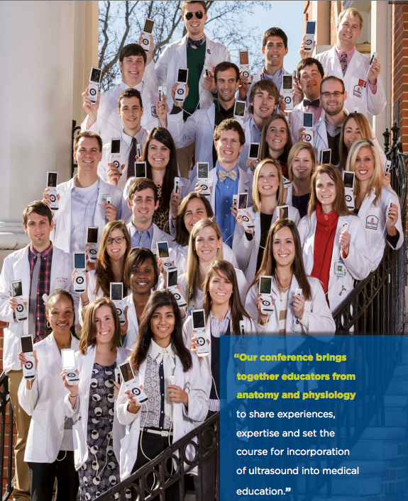
I have been to a few of the national AIUM conventions and my most recent post about it discusses one of the best days of my life, the course in NY at AIUM2013 about ultrasound integration to medical school curriculums where the experts spoke of their experiences, their advice, their ideas ….followed by a panel of medical students who shared their point of view of how it affected their education. It was amazing! Even now, when i think about it, i am inspired, and continue to be excited about how we are starting to do the same at Stanford School of Medicine. I have posted about the reactions that Ultrafest (a free medical student workshop in California) brought to everyone, including what UC Irvine’s Dean Clayman stated about it all. It was quite honest and encouraging. All of this, brings me to this message that i keep reading over and over again. Is it because 2013 is the Year of Ultrasound? Well, likely so, but for that reason and so many more, I just cant stop reading it – please read it below, especially the end when the link to the Ultrasound in Med Ed portal is introduced.

Screen Shot 2013-07-25 at 6.00.35 PM

Screen Shot 2013-07-25 at 6.00.23 PM

July 25, 2013

Dear Colleagues:

I believe there is no more important issue facing ultrasound than its incorporation into undergraduate medical schools’ curricula. Many of you saw the visionary presentation of Dean Richard Hoppmann, MD, at the 2012 AIUM Annual Convention in Phoenix, Arizona. Here at New York University School of Medicine, Uche Blackstock, MD, RDMS, an emergency department physician, is developing a multidisciplinary collaborative integrated preclerkship and clerkship curriculum. Consider the following:

It’s another typically busy day at the medical center. A critical care fellow supervises a senior resident placing ultrasound-guided central vascular access in a hypotensive septic elderly patient in the medical intensive care unit. In the outpatient surgery suite, an anesthesiologist prepares a patient for rotator cuff surgery by performing an ultrasound-guided interscalene brachial plexus block for regional anesthesia. In the echocardiography lab, a cardiologist assesses a patient admitted the night before with a non-ST segment elevation myocardial infarction for wall motion abnormalities and cardiac function. An obstetrics and gynecology attending performs a pelvic ultrasound examination on a young woman being ruled out for an ectopic pregnancy, appreciates an intrauterine pregnancy, and discharges the patient home safely.

Over the last 20 years, ultrasound performed at the bedside, by clinicians, has revolutionized the way medicine is practiced. In these diverse cases, the use of ultrasound was critical in providing patients with effective and quality clinical care. Although currently being used for diagnosis, management, and procedural guidance by physicians in numerous and diverse specialties, a significant gap currently exists between what medical students are being taught and how they are expected to practice on completion of their training. Future physicians will be expected to be familiar with the use of ultrasound in their clinical practice, regardless of specialty. At this time, medical educators have a unique and timely opportunity to use ultrasound at the bedside as an innovative teaching modality in the undergraduate medical curriculum.

Handheld ultrasound will transform how medical students are taught in the preclinical curriculum as well. Students will never experience learning medicine the same way. They will be able to scan a live model and appreciate the gallbladder as its lies within the main lobar fissure of the liver. They will have a deeper understanding of the cardiac cycle by viewing the diastolic and systolic phases of a live beating heart. During clerkships, students will learn how bedside ultrasound can be used to make important diagnoses and to allow them to safely perform critical procedures. Bedside ultrasound as a teaching tool will enhance what students have learned traditionally and help reinforce important concepts.

An integrated ultrasound curriculum will require a multidisciplinary collaborative effort by a medical school faculty. This approach will ensure that students receive comprehensive exposure to ultrasound from all perspectives. Medical educators are responsible for ensuring students are well equipped for future clinical practice. Ultrasound, as I see it, will have a dual role in undergraduate medical education. First, it can and should be incorporated into preclinical learning to teach anatomy, physiology, and pathology. Second, there is almost no clinical clerkship, as outlined by the scenarios above, that does not already or else will soon utilize bedside ultrasound. The future is now.

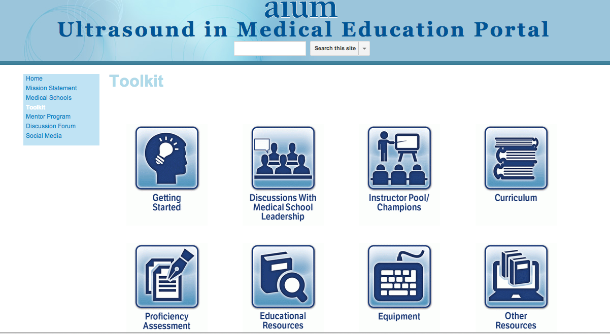
The AIUM’s Ultrasound in Medical Education Interest Group, chaired by David P. Bahner, MD, RDMS, has developed an outstanding online portal to assist those with an interest in integrating ultrasound into medical school curricula. This one-stop clearinghouse includes a mentor program; educational information from multiple organizations; and a tool kit that features curriculum examples, links to online lectures, sample proficiency assessments, tips on discussions with medical school leadership, instructor pools, equipment, and more. We encourage you to explore the Ultrasound in Medical Education Portal. If you know of additional resources that should be included, e-mail MedEd@aium.org.

In closing,

AIUM membership for students, residents, and fellows allows these individuals access to helpful ultrasound-related resources and the opportunity to network with experts in medical ultrasound–an excellent complement to the clinical training they receive. This membership category is $25 and offers students the full array of member benefits. The students of today are the future leaders of the AIUM. I hope you will share this opportunitywith those with whom you are in contact.

Sincerely,

Steven R. Goldstein, MD
AIUM President

SonoCase: Renal Ultrasound for Renal Colic: a cost/benefit analysis? by @EPMonthly #FOAMed

Once again, Drs. Teresa Wu and Brady Pregerson do an excellent job in highlighting a case in EP Monthly (and a topic that I am so incredibly passionate about – not only because of the benefit to the patient, the minimizing of CT scans/radiation, and the time spent in its work up – but also in health care cost and expediting diagnosis and management.) What am I talking about? Well, RENAL ULTRASOUND for RENAL COLIC. Yeah, I know, it sounds obvious. But, I heard of a patient the other day (again!) who had a known history of kidney stones, who had the same pain as her prior kidney stone flank pain, who begged to not have yet another CT scan done since it would have been her 13th for this at the age of 40. I highlighted this topic and other studies on it in a prior post, and AIUM posted a sound judgment series written by Drs. Chris Moore and Leslie Scoutt on this topic too.

So, let’s talk about TWu and Brady’s addition to the mix. Of course, they always start off their case with humor, yet reality, by saying : “I have to do a cost-benefit analysis of the situation,” your eager intern replies. It’s the end of the academic year and you are forcing your soon-to-be R2s to become more autonomous and confident in their management plans. You are amazed at the various answers you now get when you ask the simple question, “What do you want to do?” You ask your intern to summarize the case for you. He just finished evaluating a 21-year-old male who presented to your ED with back pain. The patient states that his “back is killing him” and he thinks he strained his muscles working out too hard at the gym last week. He just started doing CrossFit and he’s worried that he overdid it. The patient notes that the pain is 10/10 and that he has had minimal relief with his friend’s Vicodin. He’s tried icing his back and even sat in the hot tub all weekend per his friend’s recommendation. Nothing is working so his friend told him to come into the ED to get a prescription for something “stronger.”……

“Your question about whether or not this young 21 year old needs any imaging is giving him pause. “I think the cost of the imaging and the risk of radiation are too high. I don’t think there’s much benefit to keeping the patient here any longer. Plus I don’t know what we’d be looking for,” he replies. You are happy with your intern’s logic and pop into the room to see the patient. Within seconds, you realize that Vicodin and a hot tub probably won’t fix this patient’s pain. The patient is sitting hunched over on the stretcher rocking back and forth in pain. He has no appreciable tenderness to palpation over any of his back muscles, and there is no asymmetry or tightness on your exam. You are unable to reproduce or worsen his symptoms with testing his range of motion, but he is definitely rubbing his right lower back to try to ease his pain. You walk out of the patient’s room and grab your intern and the ultrasound machine. As you head back towards the patient’s room, you pimp your intern on the other more serious causes of low back pain. Acknowledging that you have the ultrasound machine in tow, your clever intern starts rattling off the diagnoses that can be easily made with bedside ultrasound. AAA, atypical appendicitis, cholecystitis, nephrolithiasis, abscess, etc. Since the patient is sitting upright and hunched over in pain, your intern decides to start his scan with a view of the right flank….”

Screen Shot 2013-07-17 at 2.37.30 PM

BAM! oh yeah – do you see it? Weren’t expecting that? Funny what happens when you look, right? You must read about their findings and the pearls and pitfalls of renal ultrasound – go here for the true meat of the article.

SonoTips & Tricks: The FAST scan: The Cardiac views #FOAMed

Finishing the ultrasound QA sessions that we do every week at Stanford, I was reminded about how bedside ultrasound is a tool that helps when resources were limited. If you hadn’t heard, there was an Asiana Airlines plane crash at San Francisco International Airport with over 180 patients requiring medical care, 55+ of which came to Stanford. Luckily, we just added 4 new SonoSite EDGE ultrasound machines to our 4 MTurbos and 3 GE Vscan systems the week before – and they sure were used well! The FAST scan was used as a screening tool and to help prioritize those who would go to the CT scanner. Once, again, it is important to know how to do it and do it well.  Our latest insert in the ACEP Ultrasound Section newsletter is below – on the FAST scan – the Cardiac sections. The prior entry was on the FAST scan: The Upper Quadrants ( go here. ) – And Ultrasound Podcast recorded with Cliff Reid about it this week too!

I’ll start with what I’ve said before: “2013 is the YEAR OF ULTRASOUND – and for good reason – there are only a few tools that give us such immediate information that can save a life. The ACEP US Section is the go-to site for everything you want to know about starting an US program, credentialing in ultrasound, the policies and politics, and is the home of SonoGuide – an amazing educational resource for bedside ultrasound, and the EMSONO: Ultrasound Test. It is also where we add our entries for their newsletter that goes over tips and tricks, cases, and all things ultrasound in the news. We recently wrote an article for the ACEP Ultrasound Section Newsletter – which is available for all members of the ACEP US Section – and I highly recommend becoming a member – it’s totally worth it.”

It was a TRUE pleasure to record a podcast recently with Dr. Scott Weingart (aka, my hero) on EMCrit (twitter: EMCrit), and writing this article with our ultrasound fellow, Dr. Viveta Lobo, describes some of what was spoken about.

By Viveta Lobo, MD and Laleh Gharahbaghian, MD, FACEP

As discussed in our last entry, the FAST exam is undoubtedly the most widely used bedside ultrasound application used in emergency medicine. Its incorporation in the ATLS revised protocol, the RUSH exam, and several other published protocols, makes it an invaluable screening tool for intra abdominal injury causing hemoperitoneum, cardiac injury with pericardial effusion, and unexplained hypotension.

We will continue our discussion of the FAST scan by reviewing the cardiac views, and relay some tips and tricks for each. Refer to the previous newsletter for tips onscanning the right upper quadrant (RUQ) and left upper quadrant (LUQ).

The Cardiac Views:

The traditional cardiac view obtained as part of the FAST exam is the subxiphoid view. The main focus of this view in the FAST exam is to evaluate for evidence of cardiac injury by evaluating for pericardial effusion and/or cardiac tamponade. The phased array probe is placed in the subxiphoid space medially, applying pressure to go under the xiphoid process and flattening out the probe while aiming caudally.

Tips for the Subxiphoid View: 

TT1 
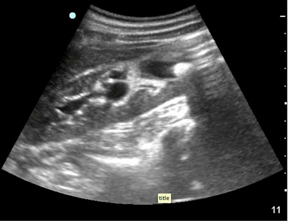
1. Use your liver as an acoustic window. 
TT2
Sound waves will travel through liver to the heart, allowing you to visualize the heart. Often one can even place the probe slightly to the right of the xiphoid process, to allow for better liver visualization, and then adjust your depth to be able to look past the liver to the heart. Without the liver in view, gas scatter will affect your image acquisition.


2. Visualize both the inferior and superior pericardial borders, to completely evaluate for pericardial effusion or, rarely, loculated pericardial effusions. It is possible for one area to have pericardial effusion and not the other. Click Here for a Video.

3. Have the patient take a deep breath and hold it. When you notice that the heart is far from the probe, and you find yourself adjusting your depth to more than 20cm, having your patient take a deep breath will lower the heart closer to the probe, improving visualization. Click Here for a Video.

TT3

Despite the subxiphoid view being the traditional view for the FAST exam, the parasternal long view is becoming more of the ‘go-to’ window to evaluate for pericardial effusion. This may be due to several very relevant clinical factors: You simply cannot get a good subxiphoid view. An injury, foreign body, or abdominal pain does not allow for subxiphoid probe placement/pressure. Or you can differentiate pericardial fluid from pleural fluid in the parasternal long view

Tips for Parasternal Long View:
As far as patient positioning, if you’ve already evaluated the RUQ and LUQ (so as to not affect free fluid evaluation) and the patient is able to turn into a left lateral decubitus position, it will help bring the heart closer to the chest wall for visualization. This can be difficult, or impossible, in trauma patients, so the below tips may help:

TT4 1. Start high and start medial – Place your phased array probe just next to the sternum, starting just under the clavicle. If you don’t see the heart there, slide down a rib space, and fan through that space to find the heart. Continue sliding down rib spaces, until you find it.

2. Slowly change the angle of your probe (up and down) when you’re assessing each rib space as described above. ‘Slowly’ is the key word here. If you’re angling downward too much in a rib space and see the PSL heart, you may need to just slide down a rib space. If that makes the image worse, slide back up.

3. Slowly rotate your probe while keeping the angle described above (clockwise/counterclockwise depending on whether you use the right shoulder or the left hip to direct your probe marker). Rotate until you visualize the longitudinal view of the left side of the heart.

4. Slide your probe medially/laterally only if you need to in order to center the aortic and mitral valves on your screen.

5. Ensure adequate depth in order to distinguish a left sided pleural effusion from a pericardial effusion. This will allow visualization of the descending thoracic aorta seen in its transverse view just deep to the heart, which is your landmark in differentiating pleural effusion from pericardial effusion. Pleural effusion will travel posterior to the aorta while pericardial effusion will travel anterior to it (and possibly circumferentially around the heart).TT5
TT6

TT7Lastly, it can be very difficult in both subxiphoid and parasternal long views to differentiate epicardial fat pad from pericardial effusion. One tip: epicardial fat is seen anteriorly and has echogenicity within it, while pericardial effusion is seen posteriorly or inferiorly and is anechoic, but can travel anteriorly if large enough to become a circumferential pericardial fluid collection. Despite this tip, clinical correlation is needed.

Look out for Part 3 of the FAST Exam: The Pelvis, in the next newsletter. Until next time, happy scanning!

For a set of links to online education in bedside ultrasound, go here. Another post on Social Media in EM Ultrasound and the amazing tools out there to learn it for free, go here.”

References
1. Ma OJ, Mateer JR, Ogata M, et al. Prospective analysis of a rapid trauma ultrasound examination performed by emergency physicians. J Trauma. 1995; 38:879-85.
2. Wherrett LJ, Boulanger BR, McLellan BA, et al. Hypotension after blunt abdominal trauma: the role of emergent abdominal sonography in surgical triage. J Trauma. 1996;41:815-20.
3. Schiavone WA, Ghumrawi BK, Catalano DR, et al. The use of echocardiography in the emergency management of nonpenetraing traumatic cardiac rupture. Ann Emerg Med. 1991;20:1248-50.
4. Rozycki GS, Feliciano DV, Ochsner MG, et al. The role of ultrasound in patients with possible penetrating cardiac wounds: a prospective multicenter study. J Trauma. 1999;46:543-52.


SonoCase: Pregnant pt with 1st trimester vaginal bleeding/pelvic pain in @EPMonthly #FOAMed

Drs. Teresa Wu and Brady Pregerson bring another engaging discussion to the great question: Should someone with a prior vaginal ultrasound for pregnancy evaluation get another one with repeat visits to the emergency department? Well, as they will describe, it may not be needed, but it sure does help patient satisfaction (and especially relief if they are concerned about their baby). So, if you do, it is all about your ability to interpret the images correctly. They identify some great vaginal/pelvic ultrasound pearls and pitfalls to keep in mind in the end of the following case:

“There are twenty-eight patients in the waiting room with the longest waiting 4 hours. The queue for CT scans is over 2 hours and the one for ultrasounds is even longer; a staggering 4 hours, plus another hour to get results. Lots of people are frustrated. Your next two patients are both pregnant females in their first trimester with vaginal bleeding. As you perform your H & P, you encounter more similarities between the two. Both have midline crampy pain like a period, with no fever, no vomiting, and no syncope. Both recently had ultrasounds done, one in your ED 3 days ago, and one with her obstetrician four days ago. You know why they are here. One reason – they want to see if their baby still has a heartbeat. You also know that repeating the ultrasound is not really medically indicated using the strict sense of the word. Sure it’s reasonable, even customary, but will it change management tonight? Can’t they just see their OB tomorrow? Is it really the right way to practice medicine to clog up your department even worse while simultaneously adding one more straw to the camel carrying the national healthcare budget? Who are you going to listen to? Press and Ganey? Barack Obama? Your conscience? What will the parents think and how will they react if you tell them, “Sorry, we can’t do an ultrasound tonight. You have to go home and make an appointment tomorrow to see your doctor.”?

The following ultrasound images are obtained in each patient:

Screen Shot 2013-07-03 at 6.39.47 PM Screen Shot 2013-07-03 at 6.40.19 PM

Do you know how to interpret them? Read more on vaginal ultrasound and their great pearls and pitfalls here.

Great pearls to keep in mind:

gestational sac only – early intrauterine pregnancy (IUP) or pseudosac of an ectopic pregnancy

gestational sac with yolks sac or fetal pole – early IUP

gestational sac with fetal pole and cardiac activity – LIVE IUP

For a review on the beta hcg (and if we can /should use ti anymore) and early pregnancy evaluation with ultrasound, go here.

SonoStudies: Thoracic Ultrasound for Pulmonary Embolism #FOAMed

Thoracic ultrasound is one of the most highly changing and advancing applications of bedside ultrasound, and the research that has been published on the utility of this application for our patients cannot be ignored. It can aid (and is better than chest Xray) in pneumothorax evaluation, pleural effusion assessment (only need 15cc of fluid to see it on ultrasound!), pneumonia evaluation, and pulmonary edema assessment. See prior posts here, here and here with literature referenced to read about all of that – trust me it’s worth it!). Of course, if you add cardiac echo to your evaluation for acute pulmonary embolism, the studies suggest it helps to look for McConnells sign and RV dilation and strain (which is a bad prognostic indicator for PE). Recently, there was a case report published in J of EM of a PE-in-transit diagnosed by bedside echo, leading to expedited care and ability to know the cause of suden cardiac arrest in a patient. For a clip of what it may look like for a “mobile mass” seen in RA, click here. Another study in J of EM was done concluding that ED bedside ultrasound echo results  predicted PE adverse outcomes.

Seeing RV dilation/strain can help but are seen mainly when the patient is hemodynamically unstable. Could thoracic ultrasound identify subsegmental pulmonary embolism in patients who are not hemodynamically unstable? Interesting question and I truly hope so…

So, if that wasnt enough, now it can help with pulmonary embolism evaluation??? What?! That is great and i hope that this teaser of a study below can be repeated and found to be valid. It would be great. Now, there have been a few others, like a meta-analysis showing that thoracic ultrasound should not be ignored when suspecting PE, a review of chest ultrasound for pulmonary diseases showing its utility, and a case report and review by the Italians (who are huge researchers in thoracic ultrasound where I listen to pretty much everything they say about it).

This recent study in Annals of Thoracic Medicine, physicians in Turkey evaluate the use of bedside ultrasound for the evaluation of pulmonary embolism. The abstract is below:

“OBJECTIVES: The diagnosis of pulmonary embolism (PE) is still a problem especially at emergency units. The purpose of study was to determine the diagnostic accuracy of thoracic ultrasonography (TUS) in patients with PE.

METHODS: In this prospective study, 50 patients with suspected PE were evaluated in Department of Pulmonary Diseases of a Training and Reasearch Hospital between January 2010 and July 2011. At the begining, TUS was performed by a chest physician, subsequently for definitive diagnosis computed tomography pulmonary angiography were performed in all cases as a reference method. Other diagnostic procedures were examination of serum d-dimer levels, echocardiography, and venous doppler ultrasonography of the legs. Both chest physician and radiologist were blinded to the results of other diagnostic method. Diagnosis of PE was suggested if at least one typical pleural-based/subpleural wedge-shaped or round hypoechoic lesion with or without pleural effusion was reported by TUS. Presence of pure pleural effusion or normal sonographic findings were accepted as negative TUS for PE.

RESULTS: PE was diagnosed in 30 patients. It was shown that TUS was true positive in 27 patients and false positive in eight and true negative in 12 and false negative in three. Sensitivity, specificity, positive predictive value, negative predictive value, and diagnostic accuracy of TUS in diagnosis of PE for clinically suspected patients were 90%, 60%, 77.1%, 80%, and 78%, respectively.

CONCLUSIONS: TUS with a high sensitivity and diagnostic accuracy, is a noninvasive, widely available, cost-effective method which can be rapidly performed. A negative TUS study cannot rule out PE with certainty, but positive TUS findings with moderate/high suspicion for PE may prove a valuable tool in diagnosis of PE at bedside especially at emergency setting, for critically ill and immobile patients, facilitating immediate treatment decision.”

From the BLUE protocol by Lichtenstein on how to distinguish the various etiologies of shortness of breath, an algorithm was given (see below) which includes the utility of bedside ultrasound for pulmonary embolism diagnosis:

A Profile: anterior A lines bilaterally only – absence of interstitial syndrome – with lung sliding

A’ profile: A profile without lung sliding

B profile – anterior B lines bilaterally with lung sliding

B’ profile – B profile without lung sliding

A/B profile – A lines on one side and B lines present on the other side (asymmetry)

C profile – anterior consolidation (shred sign)

Normal – A profile without PLAPS

PLAPS = posterolateral alveolar and/or pleural syndrome

Thoracic US and the BLUE protocol

A good presentation on thoracic US for pulmonary embolism can be found here:

SonoGallery: Free downloadable 3D & 4D OBGYN ultrasound cases and images #FOAMed

I love free stuff, especially when it is for the purpose of education. PanoramaScan is a site that sells their images and courses, but also provides a great group of free downloadable images and cases in OBGYN ultrasound. From normal fetal anatomy to molar twin pregnancy in 3D and 4D – it has some great images that are amazing to view. This truly puts a different perspective on maternal-fetal medicine.

These free downloads come with a short and brief description of the video. It is perfect for those of us who have short attention spans!

As they state in their website: “Our mission, for the past eight years, is to successfully deliver state-of-the-art 4D ultrasound and Doppler education in maternal fetal medicine (MFM) –obstetrics and gynecology (OB GYN) through our huge library of obstetrics and gynecology ultrasound courses, ultrasound cases, ultrasound teaching filessonograms (sonography images and ultrasonography videos),ultrasound web casts, ultrasound pdf documents and ultrasound ppt presentations.”

SonoTips & Tricks: The upper quadrants of the FAST scan #FOAMed

Happy Monday everyone! I mean, Tuesday!! Ugh! Well, if you were wondering where I’ve been, or even if you didn’t notice, I’ve had a busy couple weeks. From the many shifts that was full of interesting ultrasound cases (which you know I’ll share with you soon!) to graduating another stellar group of emergency residents, credentialing them in EM Ultrasound after 3 great years of training and a competency test, and hopefully soon to hear about the amazing pick-ups and lives saved in their future careers with the use of their great clinical judgement and bedside ultrasound. Im sure you love those busy weeks as much as we do, so i thought I would post something that would be a bit of positivity and highlight a group that I believe in: ACEP Ultrasound Section.

I’ll start with what I’ve said before: “2013 is the YEAR OF ULTRASOUND – and for good reason – there are only a few tools that give us such immediate information that can save a life. The ACEP US Section is the go-to site for everything you want to know about starting an US program, credentialing in ultrasound, the policies and politics, and is the home of SonoGuide – an amazing educational resource for bedside ultrasound, and the EMSONO: Ultrasound Test. It is also where we add our entries for their newsletter that goes over tips and tricks, cases, and all things ultrasound in the news. We recently wrote an article for the ACEP Ultrasound Section Newsletter – which is available for all members of the ACEP US Section – and I highly recommend becoming a member – it’s totally worth it. To continue to entice you, i will include our latest entry below – with a few additions in the end. For a set of links to online education in bedside ultrasound, go here. And, for our last entry into the Newsletter on Social Media in EM Ultrasound and the amazing tools out there to learn it for free, go here.”

Now, let’s talk about the FAST scan. It was a TRUE pleasure to record a podcast recently with Dr. Scott Weingart (aka, my hero) on EMCrit (twitter: EMCrit), and writing this article with our ultrasound fellow, Dr. Viveta Lobo, describes some of what was spoken about.

The focused assessment with sonography in trauma, or FAST exam, is undoubtedly the most widely used bedside ultrasound application in emergency medicine. With its incorporation into the ATLS trauma protocol, the FAST exam is performed immediately after the primary survey simultaneously with other resuscitative efforts. It is also a component of the RUSH protocol for patients with unexplained shock. Trauma patients often present with multiple injuries, and significant bleeding can occur without obvious changes in vitals signs. Medical patients can present intoxicated, altered, delirious, or demented all of which will limit the physical exam. The primary purpose of the FAST exam is to rapidly detect free fluid and hemorrhage in the peritoneal, pericardial and pleural spaces. There may be difficulties in obtaining adequate views, and we hope to discuss a few pearls to minimize them.

As with all ultrasound applications, familiarity with technique and patient anatomy, knowledge of common pitfalls, practice, and appreciating technical limitations are important errors to avoid. In general, the FAST exam is not “fast” – it can take up to 3-4 minutes to perform.1 The patient should be supine (or Trendelenberg) with low ambient light, with a low frequency probe used (the phased array probe provides the additional benefit of visualizing between the ribs and getting into the subxiphoid region more easily for the cardiac view). Even with the best technique, the FAST scan will only visualize 25 cc or more of thoracic free fluid and 500cc or more of intraperitoneal free fluid.2

The Right Upper Quadrant (RUQ)

RUQ

The RUQ is the most sensitive region for free fluid in comparison to the other FAST views.3 In my view, the RUQ should be divided into 3 zones.

1. Above/Below the diaphragm,
2. Morrison’s pouch (hepato-renal recess)
3. Paracolic gutter: Around the inferior hepatic edge/inferior pole of kidney

The key is to know your landmarks, and STOP, STAY and widely FAN through each zone well, adjusting your depth as necessary to keep the area of interest centered on your screen. Click Here for a Video. Start high to stay and fan (anterior to posterior) around the diaphragm. Then, SLIDE down into another rib space, stop, stay and fan around the entire kidney. An additional rib space may be necessary to evaluate the paracolic gutter.

Tips for RUQ Diaphragm View :

The liver may be easily seen, but the diaphragm can be more difficult, especially if it’s behind a rib shadow. Have the patient take in a deep breath. This lowers the diaphragm into your view and allows visualization of the thoracic cavity for hemothorax/pleural fluid as well as sub-diaphragmatic peritoneal fluid. Visualization of the spine shadow travelling in the lower part of the screen will normally stop at the diaphragm with a mirror image artifact illustrated in the thoracic cavity.

2-TT Imagespineshadow

However, if the spine is able to been seen above the diaphragm– this is pathognomonic of pleural fluid, and also known as the “V-line.”4Click Here for a Video.

3-TT Imagevline

Tips for RUQ Morrison’s Pouch (Hepato-Renal Recess) View:

If rib shadows get in the way, using the same trick above of patient inspiration can help. There are also a few false positive “traps” here.

First, the double line sign, seen around the kidney capsule as hyperechoic double lines with hypoechoic material in between, can be mistaken for free fluid.5 However, free fluid will not be surrounded by hyperechoic lines and will not be in a contained structure.

4-TT ImageVine
Second, edge artifact from the liver/kidney interface occurs due to ultrasound physics and sound wave transmission between structures of different densities. It is seen as a dark thin line tracing off the edge of this interface extending to the bottom of the screen. Click Here for a Video. This differentiates it from free fluid, which will not extend past the liver.Click Here for a Video.

5-TT ImageRUQFFinMP

Tips for RUQ Paracolic Gutter View:

This is where free fluid can be seen first amongst all the different zones of the RUQ view.6The most important tip is to not forget to view this area. You will often have to slide your probe more inferior to obtain this view. Decrease the depth to look around the hepatic edge and inferior kidney pole, and evaluate the region with slow fanning. Click Here for a Video.

6-TT ImageParaCOlicFF

The Left Upper Quadrant (LUQ)

The LUQ is less sensitive for free fluid than the RUQ for varying reasons. First, the LUQ is opposite the side of the sonographer, which can make it technically difficult to obtain an adequate view. Also, the spleen is smaller than the liver and, thus, the acoustic window is lessened.

7-TT Imagestomachsabotage

The stomach commonly obstructs the view as well. The LUQ should also be divided into 3 zones:

1. Above/Below the diaphragm,
2. Spleno-Renal recess,
3. Paracolic gutter: Around the inferior pole of kidney

Tips for the LUQ view
In addition to the various RUQ view tips and tricks as stated above, the LUQ diaphragm view also requires tips to avoid “stomach sabotage”. There are two ways around this: oblique the probe to have the indicator angled toward the gurney and/or slide your probe to the posterior-axillary line away from the plane of the stomach.

8-TT ImageLUQFFAbove

Look out for Part 2 of FAST Tips and Tricks, in the next newsletter where we talk about maximizing your cardiac views.

For additional material, images, and cases on the E-FAST, go here.

Another great review of FAST with excellent references here.

And, of course, saving the best for last – Cliff Reid and The Ultrasound Podcast discusses how to “earn your vaginal stripes” about the EFAST – go here.

Jacob Avila of 5MinSono did a great false positive blog on FAST here.

References
1.     Boulanger BR, McLellan BA, Brenneman FD, et al. Emergent abdominal sonography as a screening test in a new diagnostic algorithm for blunt trauma.
J Trauma. Jun 1996;40(6):867-    874.
2.     Branney SW, Wolfe RE, Moore EE, et al. Quantitative sensitivity of ultrasound in detecting free intraperitoneal fluidJ Trauma. Aug 1995;39(2):375-380.
3.     Chambers JA, Pilbrow WJ. Ultrasound in abdominal trauma: an alternative to peritoneal lavageArchEmerg Med. Mar 1988;5(1):26-33.
4.     Atkinson P, Milne J, Loubani O, et al. The V-line: a sonographic aid for the confirmation of pleural fluidCrit Ultrasound J. 2012;4(1):19.
5.     Sierzenski PR, Schofer JM, Bauman MJ, et al.
The double-line sign: A false positive finding on the focused assessment with sonography for trauma (FAST) examinationJ Emerg Med. 2011;40(2):188-189.
6.     Rozycki GS, Ochsner MG, Feliciano DV, et al. Early detection of hemoperitoneum by ultrasound examination of the right upper quadrant: a multicenter study.
J Trauma. Nov 1998;45 (5):878-883.

SonoStudy: Meta-analysis: History & Physical exam with Ultrasound for extremity fractures #FOAMed

I keep thinking about this study published in the Jan 2013 issue of Academic Emerg Med by Dr. Nikita Joshi et al…. for a few reasons… so I thought i would highlight it on SonoSpot and spark some discussion to get your thoughts too. (Get full article here). First off, it’s about a condition that I see in the emergency department on every shift, so it’s incredibly relevant. And, it involves imaging, specifically ultrasound, and how it can benefit the patients with this problem from cost savings to quicker diagnoses and treatment. Finally, the results actually surprised me. Not because ultrasound seemed to be just as good as radiographs, but that they weren’t better. But, I should say that it was a meta-analysis and quite difficult to compare and the study subjects in the meta-analysis all had radiograph proven fractures, and I wonder what would have happened if the xrays were negative but the bedside ultrasound was positive, proven by a gold standard, like CT scan???….  Who am I kidding?! That would involve too much cost, radiation, and time in the emergency department….. Oh wait, I get it….I guess I understand the importance of this study now. There have been quite a few studies on the topic in the last couple years – go here, here, here, here, and here – which makes it really exciting.

The authors start by stating that radiographs do miss fractures:

“The typical work-up of the injured patient generally involves a medical provider obtaining a history and physical examination, often followed by radiologic imaging. However, many times the radiologic imaging may be negative or inconclusive, which calls to question whether the imaging contributed to the management or outcome of the patient. Studies have shown that often the imaging obtained is unnecessary and results in radiation exposure to patients and increased ED wait times.[2]….There’s a low rate of positive radiography when assessing for fractures as evidenced by a retrospective review by Bentohami et al.,[3] in which only 50% of upper extremity x-rays showed fractures, and another study by Heyworth,[4] which showed 15% of patients with ankle injuries had documented fractures on x-ray. In the study by Stiell et al.,[2] patients with ankle injuries had midfoot fracture rates of 4.3%, and 9.3% had malleolar fractures. Therefore, 50% to 95% of extremity x-rays can be avoided without missing fractures.”

Ok, so we know this. Xrays arent great, so why get them? If you think the fracture would need reduction due to a displacement, then ok. But, wouldnt that be possible by physical exam as a deformed extremity so that you’d know to Xray that one? If the extremity is not deformed, but tender and swollen, why not just splint? Isnt that what you would do anyway if the xray was negative due to a high clinical concern for “occult fracture”?

The authors then follow this up with one of my favorite paragraphs on the topic:

“Bedside US has the potential benefits of reducing radiation exposure, costs, and pain, while potentially improving ED patient throughput and satisfaction. This reflects on the original purpose of developing CDRs for extremity fractures. Use of bedside US can help triage patients during a busy ED shift by quickly assessing for the presence of fracture as an adjunct to the normal history and physical examination. It can also aid nurses and physicians who may require more resources for reduction of a fracture.[11] EPs have become more adept at fracture diagnosis through independent review of US and radiographic imaging, and many researchers have examined the ability of EPs to obtain US imaging and diagnose fracture.[12, 13] Additionally, bedside US has excellent diagnostic test characteristics when performed by EPs compared to radiologists in the diagnostic evaluation for soft tissue infections,[14] cholecystitis,[15] pneumothorax,[16] or ruling out ectopic pregnancy.[17]

Love it. See the abstract below and read the entire article to see their limitations and methodology here.

Objectives

Understanding history, physical examination, and ultrasonography (US) to diagnose extremity fractures compared with radiography has potential benefits of decreasing radiation exposure, costs, and pain and improving emergency department (ED) resource management and triage time.

Methods

The authors performed two electronic searches using PubMed and EMBASE databases for studies published between 1965 to 2012 using a strategy based on the inclusion of any patient presenting with extremity injuries suspicious for fracture who had history and physical examination and a separate search for US performed by an emergency physician (EP) with subsequent radiography. The primary outcome was operating characteristics of ED history, physical examination, and US in diagnosing radiologically proven extremity fractures. The methodologic quality of the studies was assessed using the quality assessment of studies of diagnostic accuracy tool (QUADAS-2).

Results

Nine studies met the inclusion criteria for history and physical examination, while eight studies met the inclusion criteria for US. There was significant heterogeneity in the studies that prevented data pooling. Data were organized into subgroups based on anatomic fracture locations, but heterogeneity within the subgroups also prevented data pooling. The prevalence of fracture varied among the studies from 22% to 70%. Upper extremity physical examination tests have positive likelihood ratios (LRs) ranging from 1.2 to infinity and negative LRs ranging from 0 to 0.8. US sensitivities varied between 85% and 100%, specificities varied between 73% and 100%, positive LRs varied between 3.2 and 56.1, and negative LRs varied between 0 and 0.2.

Conclusions

Compared with radiography, EP US is an accurate diagnostic test to rule in or rule out extremity fractures. The diagnostic accuracy for history and physical examination are inconclusive. Future research is needed to understand the accuracy of ED US when combined with history and physical examination for upper and lower extremity fractures.

Nice job Nikita!

UltrasoundPodcast recently did a podcast on Distal radius fractures.

A great video of distal radius fractures can be seen here:

In case you’re curious about how easy it is to visualize a fracture by ultrasound, see image below. That bright white line is bone, and that break is …a break.

Screen Shot 2013-06-06 at 4.54.00 PM

SonoMedStudent: Ultrasound for Anatomy & Physiology – the lectures & the conference #FOAMed

The First Conference on Ultrasound in Anatomy and Physiology Education took place in March 2013. It was coordinated by a guru to medical student ultrasound education, Dr. Richard Hoppmann (a Dean and a proponent of US in medical education), with some of his good friends in ultrasound education, including one of my favorites, Dr. Michael Blaivas, an emergency physician that was one of the Godfather’s to bedside ultrasound and proving through his insane number of research studies that emergency physician (and others) can and should be performing bedside ultrasound for their patients.

It’s exciting, it’s relevant, and it matters. Doesn’t that feel good to your medical education?! Of course it does! What is even better, is that the lectures can all be found online for FREE here. Thank you Dean Hoppmann, and looking forward to the Second Conference coming in September. Sign up now!

To read more on Ultrasound in Medical Education and insights from the best of the best at AIUM and more, go here.

Screen Shot 2013-06-03 at 2.34.05 PM

Screen Shot 2013-06-03 at 2.34.18 PM

SonoStudy & Tutorial: Factors in testicular torsion diagnosis & treatment #FOAMed

Got to love the Canadians! This topic is also the first case that I posted in SonoSpot’s 1 year history, which reviews technique and an interesting case that baffled us yet becoming more clear with ultrasound (imagine that!). There have also been other case reports that I highlighted speaking about scrotal injuries. In this study, published in AJR, the authors set out to evaluate ultrasound accuracy, findings, and clinical predictors in pediatric testicular torsion. What factors correlate? Now, you could say that you dont need ultrasound and that physical exam alone will diagnose it, but interestingly, and not surprisingly, the physical exam isnt reliable and there have been other diagnoses made by ultrasound that helped rule in other causes of scrotal pain.

This study is a retrospective review, so take that into consideration when thinking about obstacles/limitations to the study, and the actual number of torsion cases was 35. But, it is interesting to note the factors they found with the torsion cases, particularly the ultrasound findings. Looks like color doppler is still good for something! See abstract below:

“OBJECTIVE. Testicular torsion is a common acute condition in boys requiring prompt accurate management. The objective of this article was to evaluate ultrasound accuracy, findings, and clinical predictors in testicular torsion in boys presenting to the Stollery pediatric emergency department with acute scrotal pain.

METHODS. Retrospective review of surgical and emergency department ultrasound records for boys from 1 month to 17 years old presenting with acute scrotal pain from 2008 to 2011 was performed. Clinical symptoms, ultrasound and surgical findings, and diagnoses were recorded. Surgical results and follow-up were used as the reference standard.

RESULTS. Of 342 patients who presented to the emergency department with acute scrotum, 35 had testicular torsion. Of 266 ultrasound examinations performed, 29 boys had torsion confirmed by surgery. The false-positive rate for ultrasound was 2.6%, and there were no false-negative findings. Mean times from presentation at the emergency department to ultrasound and surgery were 209.4 and 309.4 minutes, respectively. Of the torsed testicles, 69% were salvageable. Sensitivity, specificity, and diagnostic accuracy of ultrasound for testicular torsion were 100%, 97.9%, and 98.1%, respectively. Sonographic heterogeneity was seen in 80% of nonviable testes at surgery and 58% of patients with viable testes (p = 0.41).

Sudden-onset scrotal pain (88%), abnormal position (86%), and absent cremasteric reflex (91%) were most prevalent in torsion patients.

CONCLUSION. Color Doppler ultrasound is accurate and sensitive for diagnosis of torsion in the setting of acute scrotum. Despite heterogeneity on preoperative ultrasound, many testes were considered to be salvageable at surgery. The salvage rate of torsed testes was high.”

Among some other limitations, one limitation of this study is the number of torsion cases – I would have liked to have seen more – possibly a multi-site study is needed given the lack of high volume pediatric testicular torsion cases that come to the emergency department every year. Of course, there have been so many studies done that a meta-analysis can be written.

So, when you get that patient with acute scrotal pain, testicle in horizontal or abnormal lie, and an absent cremastreric reflex (and even after you have attempted to de-torse the testicle through the medial to lateral “opening a book” approach – right testicle counter clockwise, left testicle clockwise), place the patient’s leg in an open frog-leg position (you can use a towel under the scrotum to elevate and secure the scrotum in place if the patient tolerates it) and use your longer footprint linear probe. After examining the normal testicle in its transverse, longitudinal and coronal planes with and without color doppler to assess changes in echogenicity and arterial flow, examine the affected testicle the same way. Then, by using the longer footprint linear probe you can examine both testicles in the same view for adequate comparison ability.

Thanks to Dr. Turandot Saul for the images below:

An early ischemic testicle will be enlarged with no change in echogenicity, but a late ischemic testicle will be hypoechoic but may still have preserved structure: testicular torsion early

Also, a late torsed testicle will have abnormal echogenicity and structure: testicular torsion late

Normal testicle has normal echogenicity, normal color doppler flow within testicle:testicule normal flow

Testicle torsion will have absence of testicular flow and may get to the poibnt of hyperemia surrounding the testicle:

testicular late torsion extratestbloodflow   testiculartorsionnoflow

To read a medscape article on testicular torsion and ultrasound findings, go here.

SonoGuide has a great overview of the technique and images of testicular pathology – go here.

The Journal of Ultrasound in Medicine had a good review of the role of spectral doppler in early torsion, go here.

And, of course, Ultrasound Podcast has a great podcast on the how-to of Testicle Ultrasound part 1 and 2:

For another great pictorial review of testicular US and pathology, go here.