SonoCase: 25yo unresponsive, found down – by @KasiaHamptonMD #FOAMed #FOAMus

In case you all were unaware, Dr. Kasia Hampton is REALLY into ultrasound. She is a resident in emergency medicine and is teaching her colleagues how to use it. She has case after case of great findings, quick pick-ups, and lives saved and management changed due to that little old ultrasound machine. She even has another twitter/blog, called @tres_EUS  – a site for residents interested in ultrasound cases/leadership/research/etc. She emailed me this case that I thought was a fabulous use of ultrasound and actually shows what I harped on and on about with EMCrit on a recent podcast on FAST scans highlighted in our SonoTips and Tricks on FAST scan upper quadrants.

Enjoy!

“25 yo male was found unresponsive per bystanders. Upon EMS arrival he was noted to have multiple stab wounds to the upper extremities and chest. Initial set of vitals revealed tachycardia without hypotension. Patient was intubated at the scene “for airway protection”. Mechanically ventilated upon ED arrival with the following vitals: BP 135/90 mmHg, HR 105 BPM, respirations 16/min, SpO2 100%, T 35.8 C. GCS 3T. During secondary survey found to have one stab wound to the left anterior chest (inferior to the nipple), and second stab wound to the right posterior chest (lateral to the inferior aspect of the scapula). Additional two stab wounds to both shoulders were superficial and were no longer bleeding. No apparent abdominal (wall) injuries were noted. Abdomen was non-distended and soft.

The RUQ FAST scan:

Seek and ye shall find 3

FAST ultrasound evaluation was performed after the patient was log-rolled in both directions – first to the left and then to the right.  Subsequently the patient was taken to CT scan. He remained hemodynamically stable. Below the comparative findings of FAST vs CT scans.

IMAGING

FAST ULTRASOUND

CT

RUQ

perihepatic free fluid

perihepatic free fluid

SUBXIPHOID

no pericardial effusion

no pericardial effusion

LUQ

no free fluid

trace perisplenic free fluid

PELVIC

no free fluid

no free fluid

Given stab wound to left anterior chest with presence of free fluid in the abdomen (with hepatic and splenic injuries identified on CT), patient was taken to the operating room. Injury to pericardium itself without pericardial effusion was suspected on CT. During the surgical exploration it appeared that the stab wound to the left chest only nicked the pericardium (no blood within pericardial sac), while penetrating the left diaphragm, left lobe of the liver, stomach, spleen and pancreatic body.

This case illustrates a few important concepts:

  1. The ultimate importance of visualizing the paracolic gutter around inferior pole of the right kidney on FAST ultrasound exam;
  2. The dilemma of performing FAST scans after the patient has been log-rolled (in particular to the left side, while less important if rolled onto the right);
  3. The superiority of Secondary UltraSonographic Survey In Trauma (SUSS IT) over clinical exam for non-suspected injuries.

4 @broomedocs with love - SUSS IT OUT

In this particular case I wonder if the trace perisplenic free fluid would have been identified on FAST performed before log-rolling? Additionally, it is quite amazing how misleading was the clinical secondary survey in comparison to FAST findings and intra-operative discoveries. “

SonoCase: 72yo back pain & hypotensive – by Dr. Calvin Hwang @helixcardinal #FOAMus #FOAMed

Another great guest post! – by Dr. Calvin Hwang, aka @helixcardinal  – as well as the senior resident at Stanford/Kaiser EM program who updates the @StanfordEMRes residency twitter feed, provided an excellent case that illustrates a reason/indication to perform bedside ultrasound – especially the Echo/IVC and Aorta applications – illustrating why these applications are imperative to the RUSH protocol – along with good clinical judgement. Enjoy!

“Code 3 ringdown from EMS: 70 yo F coming in with 3 days of chest, back and abdominal pain, hypotensive with SBP in the 70s.

On arrival, patient is grimacing in pain, pale, diaphoretic.  She is otherwise healthy with no past medical history.  Just arrived from Thailand 1 week ago to visit her daughter and had been complaining of pain in her chest, back and abdomen.  Went to a primary care physician where she was noted to be hypotensive and sent to the ED.

Initial vital signs: BP 73/30, HR 110, T37.0, RR 25

Screen Shot 2013-08-22 at 6.21.37 PM

With the trusty bedside ultrasound, I immediately went to where I thought would be the diagnosis: ruptured AAA…..but…..

The abdominal aorta scan : I was shocked when I noticed it to be of normal caliber.  Nevertheless, I worked my way up the abdomen to the subxiphoid view when I saw:

Though it was atypical for the patient to be hypotensive and tachycardic, the presence of a pericardial effusion without tamponade suggested aortic dissection to me.  My attending got on the phone to prepare to transfer the patient while I contacted the radiologist to clear the CT scanner.  Though I attempted to view the descending aorta and aortic outflow tract on a more focused echo in the brief interim through a parasternal approach, I was unable to obtain good windows.  The IVC was plump and the rest of the FAST was negative.  A quick Chest XR was done:

Screen Shot 2013-08-22 at 6.21.01 PM

…..which did not show a wide mediastinum according to radiology.  The patient was whisked away to the CT scanner and within 45 minutes of ED arrival, the diagnosis of a Stanford type A aortic dissection with pericardial effusion (but not tamponade) was confirmed.  This would not have been possible without bedside ultrasound as I think most clinicians would have been falsely reassured by the normal CXR (widened mediastinum only present in 60% of aortic dissections1).

The patient was fluid resuscitated with crystalloid, her BP improved to 100/60 and HR came down to the 80s.  While awaiting transport, I attempted to place an arterial line for close BP monitoring.  However, approximately 60 minutes after ED arrival, the patient became progressively bradycardic and coded.  My institution’s cardiothoracic surgeons were already at bedside and performed a sternotomy with pericardial window.  Despite our efforts, we were never able to obtain return of spontaneous circulation and the patient was pronounced. These patients rarely make it to the ED due to how quickly they can decompensate, but if they do, quickening the diagnosis may help get them the intervention they need (clinical suspicion and appropriate use of bedside ultrasound is key), although a high mortality still exists.

  1. Aldeen A, Rosiere L.  “Focus on: Acute Aortic Dissection.” ACEP News, July 2009.

SonoCase: 32yo shortness of breath – by @Medialapproach #FOAMed

We have had some great additions of guest posts of cases where ultrasound mattered and helped with their diagnosis and treatment. Below is a case from Vince DiGiulio, an EMT and ED tech extraordinaire and more! – also known as@MedialApproach of the medialapproach.com as well as the founder of a great Google+ account on ultrasound. Read his case below and enjoy!
“In this case I was able to nail down the cause of the patient’s symptoms in 5 minutes, and I’m only an EMT whose US teaching has come entirely from online resources like SonoSpot. Here’s the story:

It’s a hot summer’s day and you are working a busy shift in the Minor Care unit of a community ED when a 31 year-old man presents with a chief-complaint of shortness of breath (SOB).

He states that he has been feeling SOB on exertion for the past 3-4 weeks, having attended the walk-in two weeks prior with the same complaint. There he was diagnosed with asthma and given an albuterol MDI, a course of PO steroids, and also a course of PO azithromycin “in case it was something more.” His symptoms had not improved so he decided to attend the ED for another opinion.

From the doorway you see a moderately overweight (5’9” 200#) Caucasian male in no acute distress. He is exhibiting a normal respiratory rate with no elevated work of breathing. His skin is warm and of normal color, but upon closer inspection you’re a bit surprised to notice he is actually moderately diaphoretic. He chalks it up to the outside temperature of 90 F, but it’s a chilly 70 F in the department and he’s been seated in bed for at least 20 minutes. “Hmmm,” you say to yourself.

Vital signs at rest are as follows: HR 115 bpm, RR 20/min, BP 122/68 mmHg, Temp 37.1 C.

On auscultation he has a bit of bi-basilar rales.

After obtaining a history, you head back to your desk to enter some orders when you see that an ECG and CXR were already performed at triage.

32yo M - SOB on Exertion x 3 wks_ECG

[http://sonocloud.org/files/photos/1373606099f1a0ab_o.jpg]

The ECG shows sinus tachycardia, left-atrial abnormality, left-axis deviation, poor R-wave progression, large S-wave in the right-precordial leads, and secondary ST and T-wave changes. This picture is consistent with left-ventricular hypertrophy.

Screen Shot 2013-12-02 at 12.58.36 PM

http://sonocloud.org/files/photos/13736928941892d4_o.jpg]
The CXR was read by radiology as “mild-to-moderate cardiomegaly, new from prior film (2 years ago), consider pericardial effusion.”

This Minor Care case is starting to get a bit more complicated and you’re beginning to wish you had seen the patient with chronic low-back pain instead. Not quite sure what to make of this patient’s presentation and afraid of backing up the whole department while you try to make a hard-sell on this young, otherwise healthy patient to cardiology, you instead grab your trusty ultrasound machine and head for the bedside. Here is what you see.

[http://sonocloud.org/watch_video.php?v=NAHA61SSH3B7]

In this apical 4-chamber view, you first notice that all four chambers are markedly dilated and hypokinetic. Looking specifically at the left-ventricle, it exhibits with severe hypokinesis throughout, maybe with a touch of apical akinesis. Looking closely at the apex of the LV also shows that there is an apical mural thrombus, often seen in patients with akinesis or severe hypokinesis of that region.

 [http://sonocloud.org/watch_video.php?v=SG794W7MBYBG]

As evidenced by the obvious blue jet in the left-atrium, this color-Doppler image of the mitral valve demonstrates significant mitral regurgitation. At formal echocardiography it was graded as “moderate, 2+ mitral regurgitation.”

32yo M Mitral Doppler

[http://sonocloud.org/files/photos/1373605496135c3c_o.jpg]

This pulsed-wave Doppler image shows monophasic flow through the mitral valve with a nearly absent A-wave. This proves that in addition to systolic dysfunction, the patient has significant diastolic dysfunction as well in a restrictive pattern.

[http://sonocloud.org/watch_video.php?v=SK85U7KAMSW1]

Here is a mid-ventricle parasternal short-axis view that further demonstrates the global hypokinesis of the left ventricle. It is also clear that the ventricle is large and dilated, but not hypertrophied. In this patient’s Cardiology echo, his ejection fraction was estimated in the range of 10-15%. I’m an inexperienced echocardiographer, but in addition to global hypokinesis I might specifically see some anterior-wall akinesis here as well.

[http://sonocloud.org/watch_video.php?v=KGUYWXGR2YG2]

This parasternal long axis view offers a final example of the patient’s global hypokinesis, along with a nice shot of the mitral valve. The aortic valve is also in view, but not clearly seen. Notably there is also no sign of pericardial effusion, often visible in this view if present.

So what’s our final impression? Summarizing all of the specific findings listed above, this patient has a dilated cardiomyopathy. While the workup and management of this patient could encompass a week’s worth of posts, here are the main take-home points from this case:

  1. Beware patients who are diaphoretic or tachycardic at rest. Afebrile and in no acute distress, it became essential to find a source of this patient’s few abnormalities on physical exam.
  2. Don’t be afraid of ultrasound in the Minor Care department. We like to talk a lot about the utility of ultrasound during a patient’s resuscitation, but it can be equally useful in an ambulatory setting as well.
  3. Bedside ultrasound expedites care. Without bedside ultrasound this patient would have been waiting around hours (or days) for a formal echo, if it was going to be performed at all.
  4. Sell! Sell! Sell! In most circumstances cardiology would have been very reluctant to come see an otherwise healthy 31 year-old patient, but in this case the bedside images provided immediate and definitive proof that the patient needed specialty care. It also probably gained us some street-cred with the cardiologist who could look at the saved images right in the department.
  5. Shoot first, ask questions later. In a case like this, there is no need to perform an extensive interpretation of your images at the bedside. From the very first view it was clear the patient had a dilated cardiomyopathy, so cardiology was immediately paged and the patient was readied for admission. During that time additional views were quickly obtained for later evaluation, but that first shot told us all we needed to know to make a disposition on the patient.
    As an ECG nerd, I liken it to reading the tracing of a patient with a profound wide-complex tachycardia. At the bedside there is rarely any need to get too fancy differentiating VT from SVT with aberrancy since the WCT algorithm is safe and effective for both, but once the patient is stabilized I can then go back and look for signs of AV-dissociation on the ECG to really prove it was VT.
    You think I noticed the apical thrombus in this patient’s AP4 view? No-way! That’s something Mike Mallin of the Ultrasound Podcast picked up for me when I shared the case with him. I didn’t even know how to read a pulsed-wave Doppler at the time I met the patient, but I knew how to capture the image at the level of the mitral valve so that I could review and learn from it later.

Anyway, thanks to bedside US (and you!) this patient ended up having his dilated cardiomyopathy recognized and promptly treated. Without these surprising images there’s a really good chance this patient would have been symptomatically treated for his SOB in the ED and then discharged back home. If anything, being able to reference these clips gave our emergency physician a very strong card to play in getting cardiology to take the case seriously.”

SonoSpot ! Now with SonoBilling info, SonoReferences list, SonoFellowship Curriculum & More! #FOAMed

Screen Shot 2013-11-25 at 3.24.52 PM

Remember when I stated that in August we will be revamping the SonoSpot site to include much more – well it took a little longer than one month, but Im very excited to show you what all that research and time has come to – A SonoSpot site that, well, looks the same, but with so much more exciting content!  – Information that will benefit not only those who want to learn bedside focused ultrasound and review cases and tutorials, but also those who want information on billing for ultrasound procedures, to review a list of references in each ultrasound application, as well as review a monthly fellowship curriculum that takes these references and adds the online sites and podcasts available to supplement that topic for each month!

Each of the headers will have drop down menus for even more content. Many topics on bedside ultrasound are found under SonoSpots when you go to sonospot.com. Oh, and those guest posts from others who have so patiently waited to have their awesome cases highlighted on SonoSpot will start this week too! Enjoy the new pages (…and they will keep getting enhanced with lecture videos throughout the year – it just gets better! )

Visit our SonoBilling and Privilege Form page – where you can see an estimate of the charges/wRVUs and LCD information that is all found publicly and incorporated into one excel sheet – scroll through it up/down/right/left to get it all in. Be warned: it is a ton of information that summarizes 1,000 pages of public pages into one sheet. Note the disclaimer. This page also has a sample Hospital Privilege form for those who want to get privileging in bedside ultrasound at their institution which is required at some places in order to bill, in addition to the list of items required for US billing as seen on the SonoBilling page.

Visit our SonoSmartphrases for EMRs – here is a sample of smartphrases that describe the documentation for each bedside limited ultrasound procedure that is being billed. The wording for each smartphrase is specifically stated due to the requirements for SonoBilling

Visit our SonoReferences pages – where we highlight the landmark and hot articles in each bedside focused ultrasound application, along with the link to the pubmed page for each. This is a page that is going to continually get updated as more studies get published. This is separate from our SonoStudies site, that go into further detail and discussion on specific studies that pick to highlight for various reasons.

Visit our SonoFellowship Curriculum pages. This is a sample of a curriculum should anyone want to do an ultrasound fellowship. It is a supplement to other educational opportunities that a fellow will get and describes the fellowship month-by-month on the reading assignments – including viewing online #FOAM resources for each topic (websites, blogs, podcasts, etc).

We will continue to optimize our Sonotutorials and SonoCases sites, which are our most popular sites for all bedside ultrasound believers in the world!

Hope you enjoy and, as always, I love any feedback or suggestions for additions to the site for our future upgrades.

SonoNews! Look out for updates on SonoSpot – Admin month is here!

Hi everyone! I am so very excited about the new SonoSpot pages that are coming this month! Some of you have messaged me asking:

“We love the SonoCases, can I provide a guest post with an interesting SonoCase?”

“Do you have any information on SonoBilling?”

“Im looking for a list of great SonoReferences to provide our US fellow, what would you suggest?”

“You haven’t posted in a week or so, are you alive?”

First off, I am very much alive! I have been busy packing and moving to my new home over the last 2 weeks and now that we are moved in (kind of) I am going to work on the SonoSpot site – – add even more SonoSpots that will answer all the questions and inquiries above!

Look out for guest posts by physicians who want to highlight their cases on SonoSpot where management changed, a quick diagnosis was made, and a life was saved! Also, we will be adding pages to our SonoSpots list, including the Stanford Ultrasound Fellowship Curriculum page (which can be used for anyone wanting to see what a sample fellowship curriculum looks like), a SonoBilling page which provides information on CPT codes and estimated charges for bedside limited ultrasound, and an updated SonoReference list for each application of bedside ultrasound. And, how could I not mention the SonoSmartphrases for the EMR…

After we are done doing some admin additions, we will continue our blogs on SonoTutorials, SonoCases, SonoStudies, Sonoworkshop pearls, SonoTips&Tricks, and more!

In the meantime, Ill still be adding interesting items on our Facebook page and will be continuing the always stimulating conversation on Twitter.

Hope you all find the additions helpful!

SonoNews! AIUM President on Ultrasound in Medical Education & the US in MedEd portal #FOAMed

When I read this message, a light shined so bright inside my little head, that I had to share it. I got the usual emails from AIUM (American Institute of Ultrasound in Medicine), a multi-specialty organization with thousands of members, who educate/study/encourage/collaborate on issues related to ultrasound in medicine. But, the email that came out today, a message from the new AIUM President, discusses with such ease and obviousness about how ultrasound should be integrated into medical school education.

Screen Shot 2013-07-25 at 5.53.42 PM

I have been to a few of the national AIUM conventions and my most recent post about it discusses one of the best days of my life, the course in NY at AIUM2013 about ultrasound integration to medical school curriculums where the experts spoke of their experiences, their advice, their ideas ….followed by a panel of medical students who shared their point of view of how it affected their education. It was amazing! Even now, when i think about it, i am inspired, and continue to be excited about how we are starting to do the same at Stanford School of Medicine. I have posted about the reactions that Ultrafest (a free medical student workshop in California) brought to everyone, including what UC Irvine’s Dean Clayman stated about it all. It was quite honest and encouraging. All of this, brings me to this message that i keep reading over and over again. Is it because 2013 is the Year of Ultrasound? Well, likely so, but for that reason and so many more, I just cant stop reading it – please read it below, especially the end when the link to the Ultrasound in Med Ed portal is introduced.

Screen Shot 2013-07-25 at 6.00.35 PM

Screen Shot 2013-07-25 at 6.00.23 PM

July 25, 2013

Dear Colleagues:

I believe there is no more important issue facing ultrasound than its incorporation into undergraduate medical schools’ curricula. Many of you saw the visionary presentation of Dean Richard Hoppmann, MD, at the 2012 AIUM Annual Convention in Phoenix, Arizona. Here at New York University School of Medicine, Uche Blackstock, MD, RDMS, an emergency department physician, is developing a multidisciplinary collaborative integrated preclerkship and clerkship curriculum. Consider the following:

It’s another typically busy day at the medical center. A critical care fellow supervises a senior resident placing ultrasound-guided central vascular access in a hypotensive septic elderly patient in the medical intensive care unit. In the outpatient surgery suite, an anesthesiologist prepares a patient for rotator cuff surgery by performing an ultrasound-guided interscalene brachial plexus block for regional anesthesia. In the echocardiography lab, a cardiologist assesses a patient admitted the night before with a non-ST segment elevation myocardial infarction for wall motion abnormalities and cardiac function. An obstetrics and gynecology attending performs a pelvic ultrasound examination on a young woman being ruled out for an ectopic pregnancy, appreciates an intrauterine pregnancy, and discharges the patient home safely.

Over the last 20 years, ultrasound performed at the bedside, by clinicians, has revolutionized the way medicine is practiced. In these diverse cases, the use of ultrasound was critical in providing patients with effective and quality clinical care. Although currently being used for diagnosis, management, and procedural guidance by physicians in numerous and diverse specialties, a significant gap currently exists between what medical students are being taught and how they are expected to practice on completion of their training. Future physicians will be expected to be familiar with the use of ultrasound in their clinical practice, regardless of specialty. At this time, medical educators have a unique and timely opportunity to use ultrasound at the bedside as an innovative teaching modality in the undergraduate medical curriculum.

Handheld ultrasound will transform how medical students are taught in the preclinical curriculum as well. Students will never experience learning medicine the same way. They will be able to scan a live model and appreciate the gallbladder as its lies within the main lobar fissure of the liver. They will have a deeper understanding of the cardiac cycle by viewing the diastolic and systolic phases of a live beating heart. During clerkships, students will learn how bedside ultrasound can be used to make important diagnoses and to allow them to safely perform critical procedures. Bedside ultrasound as a teaching tool will enhance what students have learned traditionally and help reinforce important concepts.

An integrated ultrasound curriculum will require a multidisciplinary collaborative effort by a medical school faculty. This approach will ensure that students receive comprehensive exposure to ultrasound from all perspectives. Medical educators are responsible for ensuring students are well equipped for future clinical practice. Ultrasound, as I see it, will have a dual role in undergraduate medical education. First, it can and should be incorporated into preclinical learning to teach anatomy, physiology, and pathology. Second, there is almost no clinical clerkship, as outlined by the scenarios above, that does not already or else will soon utilize bedside ultrasound. The future is now.

The AIUM’s Ultrasound in Medical Education Interest Group, chaired by David P. Bahner, MD, RDMS, has developed an outstanding online portal to assist those with an interest in integrating ultrasound into medical school curricula. This one-stop clearinghouse includes a mentor program; educational information from multiple organizations; and a tool kit that features curriculum examples, links to online lectures, sample proficiency assessments, tips on discussions with medical school leadership, instructor pools, equipment, and more. We encourage you to explore the Ultrasound in Medical Education Portal. If you know of additional resources that should be included, e-mail MedEd@aium.org.

In closing,

AIUM membership for students, residents, and fellows allows these individuals access to helpful ultrasound-related resources and the opportunity to network with experts in medical ultrasound–an excellent complement to the clinical training they receive. This membership category is $25 and offers students the full array of member benefits. The students of today are the future leaders of the AIUM. I hope you will share this opportunitywith those with whom you are in contact.

Sincerely,

Steven R. Goldstein, MD
AIUM President

SonoTips & Tricks: The FAST scan: The Cardiac views #FOAMed

Finishing the ultrasound QA sessions that we do every week at Stanford, I was reminded about how bedside ultrasound is a tool that helps when resources were limited. If you hadn’t heard, there was an Asiana Airlines plane crash at San Francisco International Airport with over 180 patients requiring medical care, 55+ of which came to Stanford. Luckily, we just added 4 new SonoSite EDGE ultrasound machines to our 4 MTurbos and 3 GE Vscan systems the week before – and they sure were used well! The FAST scan was used as a screening tool and to help prioritize those who would go to the CT scanner. Once, again, it is important to know how to do it and do it well.  Our latest insert in the ACEP Ultrasound Section newsletter is below – on the FAST scan – the Cardiac sections. The prior entry was on the FAST scan: The Upper Quadrants ( go here. ) – And Ultrasound Podcast recorded with Cliff Reid about it this week too!

I’ll start with what I’ve said before: “2013 is the YEAR OF ULTRASOUND – and for good reason – there are only a few tools that give us such immediate information that can save a life. The ACEP US Section is the go-to site for everything you want to know about starting an US program, credentialing in ultrasound, the policies and politics, and is the home of SonoGuide – an amazing educational resource for bedside ultrasound, and the EMSONO: Ultrasound Test. It is also where we add our entries for their newsletter that goes over tips and tricks, cases, and all things ultrasound in the news. We recently wrote an article for the ACEP Ultrasound Section Newsletter – which is available for all members of the ACEP US Section – and I highly recommend becoming a member – it’s totally worth it.”

It was a TRUE pleasure to record a podcast recently with Dr. Scott Weingart (aka, my hero) on EMCrit (twitter: EMCrit), and writing this article with our ultrasound fellow, Dr. Viveta Lobo, describes some of what was spoken about.

By Viveta Lobo, MD and Laleh Gharahbaghian, MD, FACEP

As discussed in our last entry, the FAST exam is undoubtedly the most widely used bedside ultrasound application used in emergency medicine. Its incorporation in the ATLS revised protocol, the RUSH exam, and several other published protocols, makes it an invaluable screening tool for intra abdominal injury causing hemoperitoneum, cardiac injury with pericardial effusion, and unexplained hypotension.

We will continue our discussion of the FAST scan by reviewing the cardiac views, and relay some tips and tricks for each. Refer to the previous newsletter for tips onscanning the right upper quadrant (RUQ) and left upper quadrant (LUQ).

The Cardiac Views:

The traditional cardiac view obtained as part of the FAST exam is the subxiphoid view. The main focus of this view in the FAST exam is to evaluate for evidence of cardiac injury by evaluating for pericardial effusion and/or cardiac tamponade. The phased array probe is placed in the subxiphoid space medially, applying pressure to go under the xiphoid process and flattening out the probe while aiming caudally.

Tips for the Subxiphoid View: 

TT1 1. Use your liver as an acoustic window. 
TT2
Sound waves will travel through liver to the heart, allowing you to visualize the heart. Often one can even place the probe slightly to the right of the xiphoid process, to allow for better liver visualization, and then adjust your depth to be able to look past the liver to the heart. Without the liver in view, gas scatter will affect your image acquisition.


2. Visualize both the inferior and superior pericardial borders, to completely evaluate for pericardial effusion or, rarely, loculated pericardial effusions. It is possible for one area to have pericardial effusion and not the other. Click Here for a Video.

3. Have the patient take a deep breath and hold it. When you notice that the heart is far from the probe, and you find yourself adjusting your depth to more than 20cm, having your patient take a deep breath will lower the heart closer to the probe, improving visualization. Click Here for a Video.

TT3

Despite the subxiphoid view being the traditional view for the FAST exam, the parasternal long view is becoming more of the ‘go-to’ window to evaluate for pericardial effusion. This may be due to several very relevant clinical factors: You simply cannot get a good subxiphoid view. An injury, foreign body, or abdominal pain does not allow for subxiphoid probe placement/pressure. Or you can differentiate pericardial fluid from pleural fluid in the parasternal long view

Tips for Parasternal Long View:
As far as patient positioning, if you’ve already evaluated the RUQ and LUQ (so as to not affect free fluid evaluation) and the patient is able to turn into a left lateral decubitus position, it will help bring the heart closer to the chest wall for visualization. This can be difficult, or impossible, in trauma patients, so the below tips may help:

TT4 1. Start high and start medial – Place your phased array probe just next to the sternum, starting just under the clavicle. If you don’t see the heart there, slide down a rib space, and fan through that space to find the heart. Continue sliding down rib spaces, until you find it.

2. Slowly change the angle of your probe (up and down) when you’re assessing each rib space as described above. ‘Slowly’ is the key word here. If you’re angling downward too much in a rib space and see the PSL heart, you may need to just slide down a rib space. If that makes the image worse, slide back up.

3. Slowly rotate your probe while keeping the angle described above (clockwise/counterclockwise depending on whether you use the right shoulder or the left hip to direct your probe marker). Rotate until you visualize the longitudinal view of the left side of the heart.

4. Slide your probe medially/laterally only if you need to in order to center the aortic and mitral valves on your screen.

5. Ensure adequate depth in order to distinguish a left sided pleural effusion from a pericardial effusion. This will allow visualization of the descending thoracic aorta seen in its transverse view just deep to the heart, which is your landmark in differentiating pleural effusion from pericardial effusion. Pleural effusion will travel posterior to the aorta while pericardial effusion will travel anterior to it (and possibly circumferentially around the heart).TT5
TT6

TT7Lastly, it can be very difficult in both subxiphoid and parasternal long views to differentiate epicardial fat pad from pericardial effusion. One tip: epicardial fat is seen anteriorly and has echogenicity within it, while pericardial effusion is seen posteriorly or inferiorly and is anechoic, but can travel anteriorly if large enough to become a circumferential pericardial fluid collection. Despite this tip, clinical correlation is needed.

Look out for Part 3 of the FAST Exam: The Pelvis, in the next newsletter. Until next time, happy scanning!

For a set of links to online education in bedside ultrasound, go here. Another post on Social Media in EM Ultrasound and the amazing tools out there to learn it for free, go here.”

References
1. Ma OJ, Mateer JR, Ogata M, et al. Prospective analysis of a rapid trauma ultrasound examination performed by emergency physicians. J Trauma. 1995; 38:879-85.
2. Wherrett LJ, Boulanger BR, McLellan BA, et al. Hypotension after blunt abdominal trauma: the role of emergent abdominal sonography in surgical triage. J Trauma. 1996;41:815-20.
3. Schiavone WA, Ghumrawi BK, Catalano DR, et al. The use of echocardiography in the emergency management of nonpenetraing traumatic cardiac rupture. Ann Emerg Med. 1991;20:1248-50.
4. Rozycki GS, Feliciano DV, Ochsner MG, et al. The role of ultrasound in patients with possible penetrating cardiac wounds: a prospective multicenter study. J Trauma. 1999;46:543-52.


SonoStudies: Thoracic Ultrasound for Pulmonary Embolism #FOAMed

Thoracic ultrasound is one of the most highly changing and advancing applications of bedside ultrasound, and the research that has been published on the utility of this application for our patients cannot be ignored. It can aid (and is better than chest Xray) in pneumothorax evaluation, pleural effusion assessment (only need 15cc of fluid to see it on ultrasound!), pneumonia evaluation, and pulmonary edema assessment. See prior posts here, here and here with literature referenced to read about all of that – trust me it’s worth it!). Of course, if you add cardiac echo to your evaluation for acute pulmonary embolism, the studies suggest it helps to look for McConnells sign and RV dilation and strain (which is a bad prognostic indicator for PE). Recently, there was a case report published in J of EM of a PE-in-transit diagnosed by bedside echo, leading to expedited care and ability to know the cause of suden cardiac arrest in a patient. For a clip of what it may look like for a “mobile mass” seen in RA, click here. Another study in J of EM was done concluding that ED bedside ultrasound echo results  predicted PE adverse outcomes.

Seeing RV dilation/strain can help but are seen mainly when the patient is hemodynamically unstable. Could thoracic ultrasound identify subsegmental pulmonary embolism in patients who are not hemodynamically unstable? Interesting question and I truly hope so…

So, if that wasnt enough, now it can help with pulmonary embolism evaluation??? What?! That is great and i hope that this teaser of a study below can be repeated and found to be valid. It would be great. Now, there have been a few others, like a meta-analysis showing that thoracic ultrasound should not be ignored when suspecting PE, a review of chest ultrasound for pulmonary diseases showing its utility, and a case report and review by the Italians (who are huge researchers in thoracic ultrasound where I listen to pretty much everything they say about it).

This recent study in Annals of Thoracic Medicine, physicians in Turkey evaluate the use of bedside ultrasound for the evaluation of pulmonary embolism. The abstract is below:

“OBJECTIVES: The diagnosis of pulmonary embolism (PE) is still a problem especially at emergency units. The purpose of study was to determine the diagnostic accuracy of thoracic ultrasonography (TUS) in patients with PE.

METHODS: In this prospective study, 50 patients with suspected PE were evaluated in Department of Pulmonary Diseases of a Training and Reasearch Hospital between January 2010 and July 2011. At the begining, TUS was performed by a chest physician, subsequently for definitive diagnosis computed tomography pulmonary angiography were performed in all cases as a reference method. Other diagnostic procedures were examination of serum d-dimer levels, echocardiography, and venous doppler ultrasonography of the legs. Both chest physician and radiologist were blinded to the results of other diagnostic method. Diagnosis of PE was suggested if at least one typical pleural-based/subpleural wedge-shaped or round hypoechoic lesion with or without pleural effusion was reported by TUS. Presence of pure pleural effusion or normal sonographic findings were accepted as negative TUS for PE.

RESULTS: PE was diagnosed in 30 patients. It was shown that TUS was true positive in 27 patients and false positive in eight and true negative in 12 and false negative in three. Sensitivity, specificity, positive predictive value, negative predictive value, and diagnostic accuracy of TUS in diagnosis of PE for clinically suspected patients were 90%, 60%, 77.1%, 80%, and 78%, respectively.

CONCLUSIONS: TUS with a high sensitivity and diagnostic accuracy, is a noninvasive, widely available, cost-effective method which can be rapidly performed. A negative TUS study cannot rule out PE with certainty, but positive TUS findings with moderate/high suspicion for PE may prove a valuable tool in diagnosis of PE at bedside especially at emergency setting, for critically ill and immobile patients, facilitating immediate treatment decision.”

From the BLUE protocol by Lichtenstein on how to distinguish the various etiologies of shortness of breath, an algorithm was given (see below) which includes the utility of bedside ultrasound for pulmonary embolism diagnosis:

A Profile: anterior A lines bilaterally only – absence of interstitial syndrome – with lung sliding

A’ profile: A profile without lung sliding

B profile – anterior B lines bilaterally with lung sliding

B’ profile – B profile without lung sliding

A/B profile – A lines on one side and B lines present on the other side (asymmetry)

C profile – anterior consolidation (shred sign)

Normal – A profile without PLAPS

PLAPS = posterolateral alveolar and/or pleural syndrome

Thoracic US and the BLUE protocol

A good presentation on thoracic US for pulmonary embolism can be found here:

SonoGallery: Free downloadable 3D & 4D OBGYN ultrasound cases and images #FOAMed

I love free stuff, especially when it is for the purpose of education. PanoramaScan is a site that sells their images and courses, but also provides a great group of free downloadable images and cases in OBGYN ultrasound. From normal fetal anatomy to molar twin pregnancy in 3D and 4D – it has some great images that are amazing to view. This truly puts a different perspective on maternal-fetal medicine.

These free downloads come with a short and brief description of the video. It is perfect for those of us who have short attention spans!

As they state in their website: “Our mission, for the past eight years, is to successfully deliver state-of-the-art 4D ultrasound and Doppler education in maternal fetal medicine (MFM) –obstetrics and gynecology (OB GYN) through our huge library of obstetrics and gynecology ultrasound courses, ultrasound cases, ultrasound teaching filessonograms (sonography images and ultrasonography videos),ultrasound web casts, ultrasound pdf documents and ultrasound ppt presentations.”

SonoTips & Tricks: The upper quadrants of the FAST scan #FOAMed

Happy Monday everyone! I mean, Tuesday!! Ugh! Well, if you were wondering where I’ve been, or even if you didn’t notice, I’ve had a busy couple weeks. From the many shifts that was full of interesting ultrasound cases (which you know I’ll share with you soon!) to graduating another stellar group of emergency residents, credentialing them in EM Ultrasound after 3 great years of training and a competency test, and hopefully soon to hear about the amazing pick-ups and lives saved in their future careers with the use of their great clinical judgement and bedside ultrasound. Im sure you love those busy weeks as much as we do, so i thought I would post something that would be a bit of positivity and highlight a group that I believe in: ACEP Ultrasound Section.

I’ll start with what I’ve said before: “2013 is the YEAR OF ULTRASOUND – and for good reason – there are only a few tools that give us such immediate information that can save a life. The ACEP US Section is the go-to site for everything you want to know about starting an US program, credentialing in ultrasound, the policies and politics, and is the home of SonoGuide – an amazing educational resource for bedside ultrasound, and the EMSONO: Ultrasound Test. It is also where we add our entries for their newsletter that goes over tips and tricks, cases, and all things ultrasound in the news. We recently wrote an article for the ACEP Ultrasound Section Newsletter – which is available for all members of the ACEP US Section – and I highly recommend becoming a member – it’s totally worth it. To continue to entice you, i will include our latest entry below – with a few additions in the end. For a set of links to online education in bedside ultrasound, go here. And, for our last entry into the Newsletter on Social Media in EM Ultrasound and the amazing tools out there to learn it for free, go here.”

Now, let’s talk about the FAST scan. It was a TRUE pleasure to record a podcast recently with Dr. Scott Weingart (aka, my hero) on EMCrit (twitter: EMCrit), and writing this article with our ultrasound fellow, Dr. Viveta Lobo, describes some of what was spoken about.

The focused assessment with sonography in trauma, or FAST exam, is undoubtedly the most widely used bedside ultrasound application in emergency medicine. With its incorporation into the ATLS trauma protocol, the FAST exam is performed immediately after the primary survey simultaneously with other resuscitative efforts. It is also a component of the RUSH protocol for patients with unexplained shock. Trauma patients often present with multiple injuries, and significant bleeding can occur without obvious changes in vitals signs. Medical patients can present intoxicated, altered, delirious, or demented all of which will limit the physical exam. The primary purpose of the FAST exam is to rapidly detect free fluid and hemorrhage in the peritoneal, pericardial and pleural spaces. There may be difficulties in obtaining adequate views, and we hope to discuss a few pearls to minimize them.

As with all ultrasound applications, familiarity with technique and patient anatomy, knowledge of common pitfalls, practice, and appreciating technical limitations are important errors to avoid. In general, the FAST exam is not “fast” – it can take up to 3-4 minutes to perform.1 The patient should be supine (or Trendelenberg) with low ambient light, with a low frequency probe used (the phased array probe provides the additional benefit of visualizing between the ribs and getting into the subxiphoid region more easily for the cardiac view). Even with the best technique, the FAST scan will only visualize 25 cc or more of thoracic free fluid and 500cc or more of intraperitoneal free fluid.2

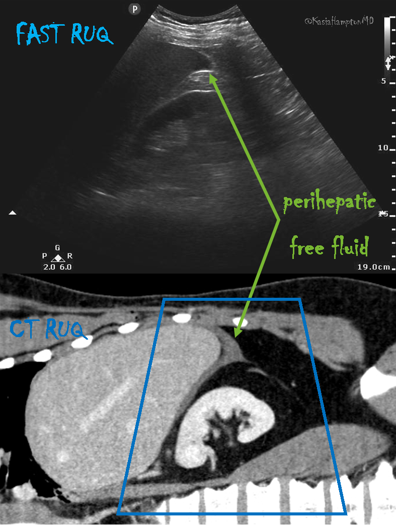
The Right Upper Quadrant (RUQ)

RUQ

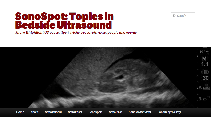
The RUQ is the most sensitive region for free fluid in comparison to the other FAST views.3 In my view, the RUQ should be divided into 3 zones.

1. Above/Below the diaphragm,
2. Morrison’s pouch (hepato-renal recess)
3. Paracolic gutter: Around the inferior hepatic edge/inferior pole of kidney

The key is to know your landmarks, and STOP, STAY and widely FAN through each zone well, adjusting your depth as necessary to keep the area of interest centered on your screen. Click Here for a Video. Start high to stay and fan (anterior to posterior) around the diaphragm. Then, SLIDE down into another rib space, stop, stay and fan around the entire kidney. An additional rib space may be necessary to evaluate the paracolic gutter.

Tips for RUQ Diaphragm View :

The liver may be easily seen, but the diaphragm can be more difficult, especially if it’s behind a rib shadow. Have the patient take in a deep breath. This lowers the diaphragm into your view and allows visualization of the thoracic cavity for hemothorax/pleural fluid as well as sub-diaphragmatic peritoneal fluid. Visualization of the spine shadow travelling in the lower part of the screen will normally stop at the diaphragm with a mirror image artifact illustrated in the thoracic cavity.

2-TT Imagespineshadow

However, if the spine is able to been seen above the diaphragm– this is pathognomonic of pleural fluid, and also known as the “V-line.”4Click Here for a Video.

3-TT Imagevline

Tips for RUQ Morrison’s Pouch (Hepato-Renal Recess) View:

If rib shadows get in the way, using the same trick above of patient inspiration can help. There are also a few false positive “traps” here.

First, the double line sign, seen around the kidney capsule as hyperechoic double lines with hypoechoic material in between, can be mistaken for free fluid.5 However, free fluid will not be surrounded by hyperechoic lines and will not be in a contained structure.

4-TT ImageVine
Second, edge artifact from the liver/kidney interface occurs due to ultrasound physics and sound wave transmission between structures of different densities. It is seen as a dark thin line tracing off the edge of this interface extending to the bottom of the screen. Click Here for a Video. This differentiates it from free fluid, which will not extend past the liver.Click Here for a Video.

5-TT ImageRUQFFinMP

Tips for RUQ Paracolic Gutter View:

This is where free fluid can be seen first amongst all the different zones of the RUQ view.6The most important tip is to not forget to view this area. You will often have to slide your probe more inferior to obtain this view. Decrease the depth to look around the hepatic edge and inferior kidney pole, and evaluate the region with slow fanning. Click Here for a Video.

6-TT ImageParaCOlicFF

The Left Upper Quadrant (LUQ)

The LUQ is less sensitive for free fluid than the RUQ for varying reasons. First, the LUQ is opposite the side of the sonographer, which can make it technically difficult to obtain an adequate view. Also, the spleen is smaller than the liver and, thus, the acoustic window is lessened.

7-TT Imagestomachsabotage

The stomach commonly obstructs the view as well. The LUQ should also be divided into 3 zones:

1. Above/Below the diaphragm,
2. Spleno-Renal recess,
3. Paracolic gutter: Around the inferior pole of kidney

Tips for the LUQ view
In addition to the various RUQ view tips and tricks as stated above, the LUQ diaphragm view also requires tips to avoid “stomach sabotage”. There are two ways around this: oblique the probe to have the indicator angled toward the gurney and/or slide your probe to the posterior-axillary line away from the plane of the stomach.

8-TT ImageLUQFFAbove

Look out for Part 2 of FAST Tips and Tricks, in the next newsletter where we talk about maximizing your cardiac views.

For additional material, images, and cases on the E-FAST, go here.

Another great review of FAST with excellent references here.

And, of course, saving the best for last – Cliff Reid and The Ultrasound Podcast discusses how to “earn your vaginal stripes” about the EFAST – go here.

Jacob Avila of 5MinSono did a great false positive blog on FAST here.

References
1.     Boulanger BR, McLellan BA, Brenneman FD, et al. Emergent abdominal sonography as a screening test in a new diagnostic algorithm for blunt trauma.
J Trauma. Jun 1996;40(6):867-    874.
2.     Branney SW, Wolfe RE, Moore EE, et al. Quantitative sensitivity of ultrasound in detecting free intraperitoneal fluidJ Trauma. Aug 1995;39(2):375-380.
3.     Chambers JA, Pilbrow WJ. Ultrasound in abdominal trauma: an alternative to peritoneal lavageArchEmerg Med. Mar 1988;5(1):26-33.
4.     Atkinson P, Milne J, Loubani O, et al. The V-line: a sonographic aid for the confirmation of pleural fluidCrit Ultrasound J. 2012;4(1):19.
5.     Sierzenski PR, Schofer JM, Bauman MJ, et al.
The double-line sign: A false positive finding on the focused assessment with sonography for trauma (FAST) examinationJ Emerg Med. 2011;40(2):188-189.
6.     Rozycki GS, Ochsner MG, Feliciano DV, et al. Early detection of hemoperitoneum by ultrasound examination of the right upper quadrant: a multicenter study.
J Trauma. Nov 1998;45 (5):878-883.