SonoStudy: Meta-analysis: History & Physical exam with Ultrasound for extremity fractures #FOAMed

I keep thinking about this study published in the Jan 2013 issue of Academic Emerg Med by Dr. Nikita Joshi et al…. for a few reasons… so I thought i would highlight it on SonoSpot and spark some discussion to get your thoughts too. (Get full article here). First off, it’s about a condition that I see in the emergency department on every shift, so it’s incredibly relevant. And, it involves imaging, specifically ultrasound, and how it can benefit the patients with this problem from cost savings to quicker diagnoses and treatment. Finally, the results actually surprised me. Not because ultrasound seemed to be just as good as radiographs, but that they weren’t better. But, I should say that it was a meta-analysis and quite difficult to compare and the study subjects in the meta-analysis all had radiograph proven fractures, and I wonder what would have happened if the xrays were negative but the bedside ultrasound was positive, proven by a gold standard, like CT scan???….  Who am I kidding?! That would involve too much cost, radiation, and time in the emergency department….. Oh wait, I get it….I guess I understand the importance of this study now. There have been quite a few studies on the topic in the last couple years – go here, here, here, here, and here – which makes it really exciting.

The authors start by stating that radiographs do miss fractures:

“The typical work-up of the injured patient generally involves a medical provider obtaining a history and physical examination, often followed by radiologic imaging. However, many times the radiologic imaging may be negative or inconclusive, which calls to question whether the imaging contributed to the management or outcome of the patient. Studies have shown that often the imaging obtained is unnecessary and results in radiation exposure to patients and increased ED wait times.[2]….There’s a low rate of positive radiography when assessing for fractures as evidenced by a retrospective review by Bentohami et al.,[3] in which only 50% of upper extremity x-rays showed fractures, and another study by Heyworth,[4] which showed 15% of patients with ankle injuries had documented fractures on x-ray. In the study by Stiell et al.,[2] patients with ankle injuries had midfoot fracture rates of 4.3%, and 9.3% had malleolar fractures. Therefore, 50% to 95% of extremity x-rays can be avoided without missing fractures.”

Ok, so we know this. Xrays arent great, so why get them? If you think the fracture would need reduction due to a displacement, then ok. But, wouldnt that be possible by physical exam as a deformed extremity so that you’d know to Xray that one? If the extremity is not deformed, but tender and swollen, why not just splint? Isnt that what you would do anyway if the xray was negative due to a high clinical concern for “occult fracture”?

The authors then follow this up with one of my favorite paragraphs on the topic:

“Bedside US has the potential benefits of reducing radiation exposure, costs, and pain, while potentially improving ED patient throughput and satisfaction. This reflects on the original purpose of developing CDRs for extremity fractures. Use of bedside US can help triage patients during a busy ED shift by quickly assessing for the presence of fracture as an adjunct to the normal history and physical examination. It can also aid nurses and physicians who may require more resources for reduction of a fracture.[11] EPs have become more adept at fracture diagnosis through independent review of US and radiographic imaging, and many researchers have examined the ability of EPs to obtain US imaging and diagnose fracture.[12, 13] Additionally, bedside US has excellent diagnostic test characteristics when performed by EPs compared to radiologists in the diagnostic evaluation for soft tissue infections,[14] cholecystitis,[15] pneumothorax,[16] or ruling out ectopic pregnancy.[17]

Love it. See the abstract below and read the entire article to see their limitations and methodology here.

Objectives

Understanding history, physical examination, and ultrasonography (US) to diagnose extremity fractures compared with radiography has potential benefits of decreasing radiation exposure, costs, and pain and improving emergency department (ED) resource management and triage time.

Methods

The authors performed two electronic searches using PubMed and EMBASE databases for studies published between 1965 to 2012 using a strategy based on the inclusion of any patient presenting with extremity injuries suspicious for fracture who had history and physical examination and a separate search for US performed by an emergency physician (EP) with subsequent radiography. The primary outcome was operating characteristics of ED history, physical examination, and US in diagnosing radiologically proven extremity fractures. The methodologic quality of the studies was assessed using the quality assessment of studies of diagnostic accuracy tool (QUADAS-2).

Results

Nine studies met the inclusion criteria for history and physical examination, while eight studies met the inclusion criteria for US. There was significant heterogeneity in the studies that prevented data pooling. Data were organized into subgroups based on anatomic fracture locations, but heterogeneity within the subgroups also prevented data pooling. The prevalence of fracture varied among the studies from 22% to 70%. Upper extremity physical examination tests have positive likelihood ratios (LRs) ranging from 1.2 to infinity and negative LRs ranging from 0 to 0.8. US sensitivities varied between 85% and 100%, specificities varied between 73% and 100%, positive LRs varied between 3.2 and 56.1, and negative LRs varied between 0 and 0.2.

Conclusions

Compared with radiography, EP US is an accurate diagnostic test to rule in or rule out extremity fractures. The diagnostic accuracy for history and physical examination are inconclusive. Future research is needed to understand the accuracy of ED US when combined with history and physical examination for upper and lower extremity fractures.

Nice job Nikita!

UltrasoundPodcast recently did a podcast on Distal radius fractures.

A great video of distal radius fractures can be seen here:

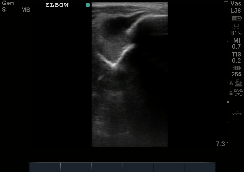
In case you’re curious about how easy it is to visualize a fracture by ultrasound, see image below. That bright white line is bone, and that break is …a break.

Screen Shot 2013-06-06 at 4.54.00 PM

SonoMedStudent: Ultrasound for Anatomy & Physiology – the lectures & the conference #FOAMed

The First Conference on Ultrasound in Anatomy and Physiology Education took place in March 2013. It was coordinated by a guru to medical student ultrasound education, Dr. Richard Hoppmann (a Dean and a proponent of US in medical education), with some of his good friends in ultrasound education, including one of my favorites, Dr. Michael Blaivas, an emergency physician that was one of the Godfather’s to bedside ultrasound and proving through his insane number of research studies that emergency physician (and others) can and should be performing bedside ultrasound for their patients.

It’s exciting, it’s relevant, and it matters. Doesn’t that feel good to your medical education?! Of course it does! What is even better, is that the lectures can all be found online for FREE here. Thank you Dean Hoppmann, and looking forward to the Second Conference coming in September. Sign up now!

To read more on Ultrasound in Medical Education and insights from the best of the best at AIUM and more, go here.

Screen Shot 2013-06-03 at 2.34.05 PM

Screen Shot 2013-06-03 at 2.34.18 PM

SonoStudy & Tutorial: Factors in testicular torsion diagnosis & treatment #FOAMed

Got to love the Canadians! This topic is also the first case that I posted in SonoSpot’s 1 year history, which reviews technique and an interesting case that baffled us yet becoming more clear with ultrasound (imagine that!). There have also been other case reports that I highlighted speaking about scrotal injuries. In this study, published in AJR, the authors set out to evaluate ultrasound accuracy, findings, and clinical predictors in pediatric testicular torsion. What factors correlate? Now, you could say that you dont need ultrasound and that physical exam alone will diagnose it, but interestingly, and not surprisingly, the physical exam isnt reliable and there have been other diagnoses made by ultrasound that helped rule in other causes of scrotal pain.

This study is a retrospective review, so take that into consideration when thinking about obstacles/limitations to the study, and the actual number of torsion cases was 35. But, it is interesting to note the factors they found with the torsion cases, particularly the ultrasound findings. Looks like color doppler is still good for something! See abstract below:

“OBJECTIVE. Testicular torsion is a common acute condition in boys requiring prompt accurate management. The objective of this article was to evaluate ultrasound accuracy, findings, and clinical predictors in testicular torsion in boys presenting to the Stollery pediatric emergency department with acute scrotal pain.

METHODS. Retrospective review of surgical and emergency department ultrasound records for boys from 1 month to 17 years old presenting with acute scrotal pain from 2008 to 2011 was performed. Clinical symptoms, ultrasound and surgical findings, and diagnoses were recorded. Surgical results and follow-up were used as the reference standard.

RESULTS. Of 342 patients who presented to the emergency department with acute scrotum, 35 had testicular torsion. Of 266 ultrasound examinations performed, 29 boys had torsion confirmed by surgery. The false-positive rate for ultrasound was 2.6%, and there were no false-negative findings. Mean times from presentation at the emergency department to ultrasound and surgery were 209.4 and 309.4 minutes, respectively. Of the torsed testicles, 69% were salvageable. Sensitivity, specificity, and diagnostic accuracy of ultrasound for testicular torsion were 100%, 97.9%, and 98.1%, respectively. Sonographic heterogeneity was seen in 80% of nonviable testes at surgery and 58% of patients with viable testes (p = 0.41).

Sudden-onset scrotal pain (88%), abnormal position (86%), and absent cremasteric reflex (91%) were most prevalent in torsion patients.

CONCLUSION. Color Doppler ultrasound is accurate and sensitive for diagnosis of torsion in the setting of acute scrotum. Despite heterogeneity on preoperative ultrasound, many testes were considered to be salvageable at surgery. The salvage rate of torsed testes was high.”

Among some other limitations, one limitation of this study is the number of torsion cases – I would have liked to have seen more – possibly a multi-site study is needed given the lack of high volume pediatric testicular torsion cases that come to the emergency department every year. Of course, there have been so many studies done that a meta-analysis can be written.

So, when you get that patient with acute scrotal pain, testicle in horizontal or abnormal lie, and an absent cremastreric reflex (and even after you have attempted to de-torse the testicle through the medial to lateral “opening a book” approach – right testicle counter clockwise, left testicle clockwise), place the patient’s leg in an open frog-leg position (you can use a towel under the scrotum to elevate and secure the scrotum in place if the patient tolerates it) and use your longer footprint linear probe. After examining the normal testicle in its transverse, longitudinal and coronal planes with and without color doppler to assess changes in echogenicity and arterial flow, examine the affected testicle the same way. Then, by using the longer footprint linear probe you can examine both testicles in the same view for adequate comparison ability.

Thanks to Dr. Turandot Saul for the images below:

An early ischemic testicle will be enlarged with no change in echogenicity, but a late ischemic testicle will be hypoechoic but may still have preserved structure: testicular torsion early

Also, a late torsed testicle will have abnormal echogenicity and structure: testicular torsion late

Normal testicle has normal echogenicity, normal color doppler flow within testicle:testicule normal flow

Testicle torsion will have absence of testicular flow and may get to the poibnt of hyperemia surrounding the testicle:

testicular late torsion extratestbloodflow   testiculartorsionnoflow

To read a medscape article on testicular torsion and ultrasound findings, go here.

SonoGuide has a great overview of the technique and images of testicular pathology – go here.

The Journal of Ultrasound in Medicine had a good review of the role of spectral doppler in early torsion, go here.

And, of course, Ultrasound Podcast has a great podcast on the how-to of Testicle Ultrasound part 1 and 2:

For another great pictorial review of testicular US and pathology, go here.

SonoStudy: 550 pts, prospective study: How good is ultrasound for traumatic pneumothorax? @westjem #FOAMed

In the march 2013 issue of Western Journal of Emergency Medicine, a study done that has been described as having generalizability, as the ultrasound scans were by many different levels of physicians, prospectively, during a trauma assessment for pneumothorax, has caused quite a bit of discussion. Mostly due to some of the limitations of the study. It is great that a prospective study with some generalizability is seen, but I wonder about the details in the methodology. They begin by discussing the importance and relevance of ultrasound for pneumothorax:

“Rapid diagnosis and treatment of traumatic pneumothorax (PTX) is important to prevent tension physiology and circulatory collapse in patients with blunt and penetrating trauma. Supine chest radiograph (CXR) is traditionally employed; however, it misses up to 50% of PTXs.1 Thoracic ultrasound (TUS) was first described in 1995 for diagnosing PTX in humans when Lichtenstein noted that the absence of comet-tail artifacts and lung sliding were associated with PTX.2 Since then ultrasound has become a validated method of examining the pleura in multiple settings. In 2011 the Eastern Association for the Surgery of Trauma gave a level 2 recommendation for the use of ultrasound to identify PTX in its practice management guidelines.3 In most studies TUS has been found to have favorable results. In Lichtenstein’s study,2 TUS had a sensitivity and negative predictive value of 100% and 96.5%, respectively, for the detection of PTX in the intensive care unit setting.4 Dulchavsky5 subsequently demonstrated that this modality has a sensitivity of 95% in the detection of PTX in patients at a Level 1 trauma center. These reports used plain radiography as the gold standard: a diagnostic modality known to be inaccurate in the detection of PTX.6 In subsequent studies using dedicated chest computed tomography (CCT) as a reference standard, sensitivities of TUS have ranged widely from 49% – 98%, while finding that it is still consistently more accurate than supine CXR.713 Studies in which TUS is performed by emergency physicians (EP) for traumatic PTX have reported even higher sensitivities ranging from 86–97% with specificities of > 99%.14 While these latter numbers are desirable, they have the potential limitation of being less applicable due to a higher skill level of the sonologists involved. The actual performance of TUS for PTX would likely vary based on the sonologist’s skill and experience. The current investigation set out to determine the test characteristics of TUS for traumatic PTX in the hands of a large heterogenous group of potential sonologists representative of typical clinicians involved in trauma care.”

The full abstract is shown below:

“Introduction:

Prior studies have reported conflicting results regarding the utility of ultrasound in the diagnosis of traumatic pneumothorax (PTX) because they have used sonologists with extensive experience. This study evaluates the characteristics of ultrasound for PTX for a large cohort of trauma and emergency physicians.

Methods:

This was a prospective, observational study on a convenience sample of patients presenting to a trauma center who had a thoracic ultrasound (TUS) evaluation for PTX performed after the Focused Assessment with Sonography for Trauma exam. Sonologists recorded their findings prior to any other diagnostic studies. The results of TUS were compared to one or more of the following: chest computed tomography, escape of air on chest tube insertion, or supine chest radiography followed by clinical observation.

Results:

There were 549 patients enrolled. The median injury severity score of the patients was 5 (inter-quartile range [IQR] 1–14); 36 different sonologists performed TUS. Forty-seven of the 549 patients had traumatic PTX, for an incidence of 9%. TUS correctly identified 27/47 patients with PTX for a sensitivity of 57% (confidence interval [CI] 42–72%). There were 3 false positive cases of TUS for a specificity of 99% (CI 98%–100%). A “wet” chest radiograph reading done in the trauma bay showed a sensitivity of 40% (CI 23–59) and a specificity of 100% (99–100).

Conclusion:

In a large heterogenous group of clinicians who typically care for trauma patients, the sonographic evaluation for pneumothorax was as accurate as supine chest radiography. Thoracic ultrasound may be helpful in the initial evaluation of patients with truncal trauma.”

So what are the limitations? They describe a few of them:

The technique: “The TUS examination consisted of the consecutive sonographic interrogation of every intercostal space between the clavicle and the diaphragm on each hemithorax. Scans were performed in the mid-clavicular line. On the left side, if cardiac motion was encountered in the mid-clavicular line, the probe was moved laterally to the left anterior axillary line and the pleura seen in the remaining intercostal spaces was evaluated until the diaphragm/spleen was encountered. To use the ribs to assist in the identification of the rib spaces and the pleura, the probe was placed in a longitudinal plane for the entire exam.” So, would this have increased their sensitivity or specificity as they include all rib spaces? Not too sure. Is this truly generalizable if the technique is different than how most perform the quick E-FAST? no. The main reason for the technique, i imagine, is to find the lung point which is far more specific for pneumothorax.

The Probe and Machine – The low frequency curvilinear probe was used on an older ultrasound system – SonoSite Titan. Could this have affecte their results? Would the increased resolution of a linear probe have helped their evaluation on the newer machines? It is possible, but by how much? who knows.

The comparison group: “Not all subjects underwent CCT and instead just had CXR and clinical observation. It is possible that some patients in this latter group had radio-occult PTX that may have been visualized on CCT leading to misclassification bias. Such a bias could result in a lower sensitivity rate for both TUS and CXR, however would likely not affect the accuracy of these tests for determining clinically significant PTX.” It is tough to have a standard and if only the chest CT group were compared, it may have had different results.

Im hoping to see more studies like this one where more generalizability is seen, and not studies done only by the experts, so that we can have a true assessment. It is best done using the technique most commonly performed (using only the second intercostal space and mid clavicular line and trying to ind the lung point if absence of lung sliding is seen) at multiple-sites, with increased power to the study, all compared to a CT as the imaging gold standard. But, i can dream, as that is quite difficult to accomplish, and the authors did a pretty nice job with what they had, got pretty good numbers of subjects – something to ponder….

For a prior post on pneumothorax and a link to the CHEST meta-analysis, go here.

For a SonoTutorial post on pneumothorax ultrasound, go here.

Another study stating that ultrasound can be used to assess post=procedure pneumothorax published in June 2013 of JUM, go here.

SonoCase: 45 yr male- flank pain & hematuria- not always a kidney stone – by Dr. Marzec et al. in @westjem #FOAMed

Western Journal of Emergency Medicine must be great proponents of bedside ultrasound! I love that journal! Of course, I am biased as I am one of the section editors, but these cases deserve mention. There have been quite a few in the March 2013 issue and this case in particular is a great review of renal ultrasound and what to lookout for in bedside ultrasound. Limited renal ultrasound typically involves an evaluation for hydronephrosis, but it is important to know what normal ultrasound anatomy looks like, as you may identify something else…. Dr. Marzec et al. at USC do a great job at discussing their case, describing their ultrasound, and giving a literature review on the finding. The case:

“A 45-year-old male with no previous medical history presented to the emergency department (ED) with 1 week of hematuria and left flank pain. The patient had noted that over the preceding 4 days his urine had progressed from a pink color to dark red. He had also experienced left flank pain that was sharp, non-radiating, and increasing in severity over the week prior to presentation. He denied a history of renal calculi, weight loss, fevers, fatigue, or abdominal masses. Upon physical examination, his vital signs included blood pressure of 157/89 mmHg, heart rate of 64 beats/min, temperature of 97.4 °F, respiratory rate of 18 breaths/min, and oxygen saturation of 99% on room air. The patient appeared comfortable. His abdomen was soft, non-tender and non-distended. The patient had left-sided costo-vertebral angle tenderness to palpation. There was frank hematuria in the urine sample at bedside. Subsequent microscopic analysis revealed > 50 red blood cells and 4–10 white blood cells. Bedside emergency ultrasound (EUS), initially performed to look for hydronephrosis, showed ….”

To read on the case, what happened, and a great review of the literature of ultrasound’s utility with this finding compared to other imaging modalities, go here.

Sono-iBook: Intro to Bedside Ultrasound – great chapters/images/videos – Volume 1& 2 in iTunes! #FOAMed

LLLLLLLLet’s get ready to UltraSoooooooouuuuuuuuund!!!! It’s what we have all been waiting for! It’s finally here! Weighing at a meager zero pounds (since it’s on the iPAD, oh yeah!), another amazing product of Drs. Mike Mallin and Matt Dawson of ultrasoundpodcast fame, and authors including experts in bedside ultrasound from around the world (and little ole’ me too). I’d like to present the SECOND volume of the Introduction of Bedside Ultrasound ! And, as Mike and Matt say it best, “If you already own Volume 1….” (which include topics in basic ultrasound applications & more filled with visual image and video clip tutorials – unlike any other “text”book that you have ever owned!) “…..this is much better.  If you don’t yet own Volume 1….they’re equal…..get them both.” – Yes, trust me, you will not be disappointed. You can also get Volume 1 on inkling chapter by chapter purchasing ability where you can read it on your iPHONE too!)Take your iPAD to the bedside, place it on the ultrasound machine, or both to help guide your ultrasound education and that of others! Volume One pics:

Screen Shot 2013-05-17 at 12.03.13 PMScreen Shot 2013-05-17 at 12.03.21 PM

Volume Two involves topics on TEE (which is an up and coming application of bedside ultrasound in cardiac arrest), MSK, Right Heart, EMS, Medical Education, Gallbladder, more Nerve Blocks (that’s where I come in..heehee :), PIV, Soft Tissue, DVT, Appy, Peds, Diastology, and much more!  364 pages of interactive content, with HOURS of video demonstrations and tutorials. – doesn’t that make you drool!?! In volume two picture: …do you know what technique that is? you will…

Screen Shot 2013-05-17 at 12.03.33 PM

To hear them speak on it, and to get a taste of perfection, go here.

Screen Shot 2013-05-17 at 12.03.24 PM and Screen Shot 2013-05-17 at 12.03.28 PM

SonoCase: 22yr old male blunt trauma to scrotum – by Dr. Cannis et al. in @westjem #FOAMed

March 2013 was a great month for ultrasound case reports and publications – especially in Western Journal of Emergency Medicine! Once again, the team from USC highlight a case where ultrasound is used at its best. As they state: “its greatest asset lies in the ability to rapidly make the diagnosis of a time-sensitive medical condition, enabling the [emergency phsyician] to mobilize resources and expedite treatment, which might otherwise be delayed. The use of [emergency] US for the evaluation of scrotal injury from blunt trauma exemplifies this point.” Isn’t it great when you include ultrasound in your examination of a patient who you will call a specialist for anyway, but to also describe the injury to them in detail, including whether there is hematoma, blood flow, or other findings – and expedite specialty care? YES! They do an excellent job in describing scrotal anatomy, the risks of missing injuries, and the findings of the case while reviewing scrotal ultrasound and the literature around it as well. This is worth the time to read it!

The case: “22-year-old male with no significant past medical history presented to the Emergency department approximately 3 hours after he was in an altercation, during which he sustained multiple blows to the head, stomach, and genital area with a large flashlight. His primary complaint was of severe testicular pain.

Physical examination revealed a calm, well-developed male in mild distress due to pain. Vital signs included a blood pressure 132/85 mmHg, heart rate of 90 beats per minute, respiratory rate 16 breaths per minute, and temperature 98.9°F. On examination of the genitals, the penis was normal. His scrotum was enlarged to approximately the size of a grapefruit, and the overlying skin was erythematous. The scrotal area was exquisitely tender to palpation, making it impossible to reliably identify or examine either testis, despite the use of parenteral opioid analgesia. A urinalysis was obtained, which was normal and notably negative for blood.” The ultrasound study showed:

Read on more, as there are more videos, and a great description of scrotal trauma and injuries with an evidence based review.


A great pictorial review of testicular ultrasound and pathology, go here.

SonoCase: 61yr old with leg swelling, chronic cough, intermittent chest pressure – by Dr. Torregrossa et al. in @westJEM #FOAMed

This case is one where if I were the doctor, my immediate response may have been hidden from the patient. Inside voice would NOT have stayed in. Wow! Dr. Torregrossa and the team at USC discuss a case published in March 2013 Western Journal of Emergency Medicine of a patient where there obviously was no bedside ultrasound performed for the duration of his symptoms. “How long was that?” you may ask… ONE YEAR! Wow! He saw his doctor (check), he got a chest Xray (check), he got an EKG (check). Good thing he finally got an ultrasound study ….

The case: “61-year-old male with a 1-year history of bilateral lower extremity swelling and a chronic cough was referred to the emergency department (ED) for an abnormal echocardiogram. The patient also reported experiencing intermittent episodes of chest pressure. He stated that he was referred from his doctor after he received a cardiac echocardiography examination that showed possible mitral valve vegetations. On review of systems, he also admitted to intermittent chest palpitations. On physical examination, his vital signs included a blood pressure of 127/75 mmHg, heart rate of 80 per minute and regular, respiratory rate of 18 per minute, pulse oximetry of 98% and temperature of 98.0°F. The rest of the physical examination was normal. An electrocardiogram demonstrated normal sinus rhythm and the chest radiograph was unremarkable. ED bedside ultrasound (EUS) showed….”

To read on the topic so that you will know some of the literature behind it – go here.

SonoCase: 32yr old with right flank pain, fever, cough – by Dr. McKaigney in @westJEM

In the March 2013 issue of Western Journal of Emergency Medicine, Dr. McKaigney highlights a case that illustrates there is more to a thoracic and right upper quadrant bedside ultrasound study than just free fluid, renal and gallbladder evaluation. You must look everywhere and appreciate when something looks abnormal. I always say, know what NORMAL looks like, because when you see something abnormal, you’ll identify at least that, then want to find out what that abnormality is by further testing.

The case: “A 35-year old male presents to the emergency department (ED) with what he describes as right-sided upper back and flank pain, which he attributes to a “cupping” procedure the day prior. The cupping procedure is an alternative medicine practice that uses local suction to theoretically stimulate blood flow and promote healing. He had no previous issues with the procedure. On further history he reported having had approximately 6 weeks of intermittent fevers, cough, anorexia and general malaise. He had seen multiple naturopathic physicians for these complaints, before an urgent care visit one week earlier. At that time, he had been started on azithromycin and doxycycline for a presumptive diagnosis of pneumonia. In the interim week he reported an improvement in his febrile symptoms and overall well-being. He was an otherwise healthy heterosexual male, without drug use or travel outside the country. He had no known sick contacts.

On physical examination his vital signs included a blood pressure of 116/75 mmHg, a heart rate of 119 beats per minute, and a respiratory rate of 20 breaths per minute. His temperature in the ED was 36.2°C. Oxygen (O2) saturation was 97% on room air. The patient was alert, and appropriate with no signs of respiratory distress. Pertinent physical findings revealed typical, non-tender cupping marks on his back. More concerning was an absence of breath sounds on the right side of the chest on auscultation. His abdomen was soft and non-tender. The remainder of the physical examination was non-contributory.

The initial diagnostic test ordered was a chest radiograph, which showed 80% opacification of the right hemithorax, consistent with pneumonia and associated parapneumonic effusion seen in Figure 1. A bedside ultrasound was subsequently performed in the ED, initially in order to examine the size of the pleural effusion in which a startling discovery was made…..”

Screen Shot 2013-05-01 at 2.28.40 PM

So, “what is that?” – you may be asking…. and what happened to the patient, what can be done about it, and what is the evidence based review on the topic? Read on as Dr. McKaigney does an excellent job in discussing it all….here

SonoCase: 72yo demented, abdominal distension -by Drs Teresa Wu/Brady Pregerson in @EPMonthly #FOAMed

Once again another great case by Drs. Teresa Wu and Brady Pregerson in EP Monthly. Whenever I read their cases, I can actually imagine myself going through the case too. This is especially true for this one, as it is a prime example of how ultrasound can get you the diagnosis immediately, and how ultrasound can be utilized in the elderly and demented nursing home patients who get sent to the emergency department for “she just doesn’t seem normal” or, in this case, “abdominal distension”. Trust me, both can actually end up with the same diagnosis. It’s also a great entry as it speaks of a procedure that all emergency physicians should know how to do – it is too easy!

The case: “72-year-old male brought in by his nursing home aide for abdominal distension. He has a history of dementia and is primarily bedridden at baseline. The patient cannot give any reliable history, but on physical exam, his otherwise thin abdomen shows obvious signs of suprapubic distension. Your intern recaps his vital signs, which include tachycardia at 120 bpm, a blood pressure of 190/86 mmHg, a respiratory rate of 20/min, and a normal temperature and O2 saturation.”…. So, the differential diagnosis? Well, you should always think of the most emergent first, like an abdominal aortic aneurysm, which can also be diagnosed by ultrasound immediately – as discussed in a prior post of another elderly patient with altered mental status. (To see more sonocase posts in evaluating the altered patient, go here). Other badness? perforated bowel, volvulus, mesenteric ischemia, hemorrhage…. Oh, the list keeps going on and on when you have an elderly patient, a demented patient, a nursing home patient – or, in this case, it was all of the above!

Whenever I am evaluating the elderly patient with abdominal complaints, I think bedside ultrasound immediately (of course, with a very low threshold for CT scan since they can have anything happen! – and let’s be honest, they aren’t the ones we think about when we talk of the radiation risks… But, healthcare bill/cost? That’s a whole other conversation…). After as best of a history and physical exam that I can get (it can be challenging when they are demented and no caregiver at the bedside! Calling the nursing home is always done but usually they are too sick or the person on the other end of the line gives limited information), I bring my ultrasound machine and explore their abdomen: FAST (which also gives you a good look at the kidneys for hydronephrosis), Aorta, Gallbladder, Bladder, Bowel, +/- Pelvic/Testicular (depending on exam). Doing that may give you the answer, as in the case highlighted above…. to find out what they found and what happened to that patient, read on here. Trust me, you’ll love it.