Unknown's avatar

About SonoSpot

US Director Emeritus, Stanford Emergency Medicine Process Improvement Director, Stanford Emergency Medicine Co-Chair, Case Review Committee, Stanford Emergency Medicine

SonoStudy: CT vs Ultrasound, Community vs Academic Ctr – kids evaluated for appendicitis

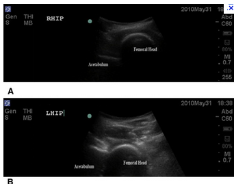
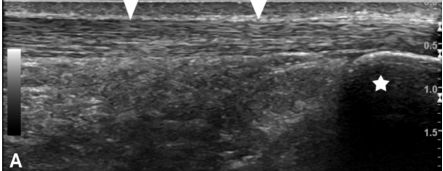
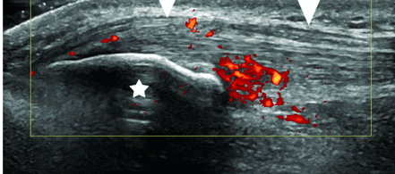
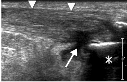
In a recent Mescape news article, a topic near and dear to my heart (and, yes, I know I have a lot of them – but decreasing radiation exposure, length of stay, and health care cost are a few), there was a study highlighted that compared community practice versus academic practice in the evaluation of children with abdominal pain that required imaging for ruling out appendicitis. It basically states that community practice do more CT scans and the results are less sensitive. Ive copied the article below, but it got me thinking…. there are quite a few factors that are different in community practice from academic practice and I wonder if they bias these results. Some community practice groups do perform published research studies, but academic centers are well known for being the research hub – does that mean they are more in tune with the talk around town? or that they are more progressive? Well, that can be argued as quite a few academic centers may seem like they are resistant to change. Also, is ultrasound available 24/7? Many community practice centers do not have access to ultrasound outside of business hours, and I know that a few academic centers are also ultrasound-openic overnight. The radiologists who read these studies may not even be in the same country as out-sourcing has become more common than ever before. Would that decrease the sensitivity? It’s hard to say, but I doubt they would be in demand if they made that many mistakes. Surgeons are more reluctant to take a patient to the operating room without a CT-proven appendicitis and emergency physicians are less likely to discharge a patient without a clear diagnosis for right lower quadrant pain. Do any of these factors play into this? Hmmmm…..well, in a prior post about a study done on ultrasound versus CT, the numbers suggest that change is needed…. somewhere along the line of the work up.

“Community hospitals are more than 4 times more likely than pediatric institutions to use radiation-exposing computed tomography (CT) scans and 80% less likely to use ultrasound for pre-appendectomy evaluations in children, study results suggest. Jacqueline M. Saito, MD, MSCI, and colleagues from Washington University School of Medicine in St. Louis, Missouri, also found that both diagnostic tools were less sensitive for appendicitis in the community hospital setting. As previously reported by Medscape Medical News, CT screening of children with abdominal pain has skyrocketed while appendicitis rates remain unchanged, adding to growing concerns regarding the link between excessive radiation exposure and cancer risk later in life. “Broadly-applicable strategies to systematically maximize diagnostic accuracy for childhood appendicitis, while minimizing ionizing radiation exposure, are urgently needed,” the authors write, noting that evaluations may be streamlined by using algorithms developed with broad validity to decrease reliance on preoperative imaging and radiation exposure while avoiding unnecessary hospital transfers, admissions, operations, and missed diagnoses. The retrospective study was published online December 24 in Pediatrics.

For the study, researchers reviewed the records of 423 children who had undergone surgery for presumed appendicitis. Preoperative imaging was performed in 93.4% of cases; final diagnoses included acute appendicitis (69.0%), perforated appendicitis (23.6%), and normal appendix (7.3%). After adjusting for age, sex, race/ethnicity, body mass index, symptom duration, and white blood cell count, researchers found that children initially evaluated at a community hospital were 4.4 times more likely to have undergone a preoperative CT scan (odds ratio [OR], 4.37; 95% confidence interval [CI], 1.70 – 11.19; P = .002) and 80% less likely to have had an ultrasound performed (OR, 0.20; 95% CI, 0.07 – 0.58; P = .003) than those at a pediatric facility. About 15.1% of children underwent both ultrasound and CT before surgery, particularly if they were girls (OR, 4.51; 95% CI, 1.47 – 13.82; P = .008) or had a lower body mass index percentile (OR, 0.98; 95% CI, 0.96 – 1.00; P = .03), longer symptom duration (OR, 1.81; 95% CI, 1.15 – 2.86; P = .01), or lower white blood cell count (OR, 0.87; 95% CI, 0.78 – 0.97; P = .01). Most children undergoing both tests had the ultrasound first (46/64, 71.9%), and normal/indeterminate results were followed up with CT (OR, 17; 95% CI, 7.7 – 37.0). Although high overall, CT scans performed at pediatric hospitals tended to be more sensitive for any appendicitis and for perforated appendicitis than those done at community hospitals (98.8% vs 93.4% [P = .07] and 75.0% vs 49.0% [ P = .045], respectively). Sensitivities were highest for older children (aged 13 – 18 years) and those not obese; insufficient numbers of underweight children were available for analysis. Accuracy of ultrasound for diagnosing appendicitis was found to be moderate in the pediatric hospital setting (weighted κ, 0.36; 95% CI, 0.24 – 0.48) and highest among older children (aged 13 – 18 years; weighted κ, 0.38; 95% CI, 0.22 – 0.54) and boys (weighted κ, 0.40; 95% CI, 0.21 – 0.55); rarity of use in community hospitals precluded any evaluation of ultrasound sensitivity in this setting. “Variation in diagnostic imaging use for pediatric appendicitis by initial evaluation location might stem from multiple factors, such as availability of imaging or the perceived need for diagnosis confirmation,” the authors comment, noting that ultrasound may be less available in community hospitals and that emergency physicians may have low risk tolerance for pediatric diagnostic errors and malpractice claims, preferring to place their confidence in CT scans.” Pediatrics. Published online December 24, 2012. Abstract

This has been talked about in further studies, as the ED length of stay can be reduced when utilizing US, instead of CT

2012 in review for SonoSpot – Thanks for the support! Happy New Year!

100 posts, 23,000 views from 119 countries!?! Wow! One year ends, another year begins. This time always comes every year when we look back on all that made us feel happy, sad, and like jumping up and down in sheer joy and excitement. Believe it or not, SonoSpot started just a meer 7 months ago, and it has been a place where we can share, highlight, and illustrate all aspects of bedside ultrasound – through our blog, twitter newsfeed, and facebook posts. With every post, comment, debate, and communication you should picture me jumping up and down because I get to do what I love – work with amazing people, educate and learn from others (and that includes all of you who post/tweet/blog/podcast/hangout/etc – thanks for making me smarter), and spread the gospel of ‘sound to the masses! It has been a true pleasure, and I thank you, from the bottom of my heart, for the support.

2012 annual report for SonoSpot. These numbers are huge to me – blows my mind, actually. Now, I know others are in the millions, as they should be –  they are incredibly amazing – they are all like mama and papa bears to me (the brown-haired step child), but starting with this kind of support makes me giggle and shake in excitement!  And, it wouldn’t be possible without you. Thank you so much! –  for reading, for sharing, for wanting to learn bedside ultrasound – and a huge thanks to all those who educate/research/share their cases/experiences with it as well. i will continue to highlight your work always.

“You complete me.” – Tom Cruise in Jerry Maguire

In just a short 7 months:

4,329 films were submitted to the 2012 Cannes Film Festival. This blog had 23,000 views in 2012. If each view were a film, this blog would power 5 Film Festivals.

In 2012, there were 100 new posts, not bad for the first year!  The busiest day of the year was August 15th with 352 views. The most popular post that day was SonoStudies: Ultrasound First for Appendicitis, the gift that keeps on giving…..

119 countries in all! Most visitors came from The United States. Australia & Canada were not far behind.

Attractions in 2012

These are the posts that got the most views in 2012.

In 2013 SonoSpot hopes to bring you even more:

– Complete SonoTutorials on all applications for bedside ultrasound

– SonoReference lists linking back to the research study

– SonoLectures – a 5 minute review of each application

– Advance and enhance the SonoMedStudent and SonoGlobalHealth sections

– and more SonoCases, SonoStudies, SonoTips&Tricks that show how bedside ultrasound can quicken diagnoses, change management, and save a life.

SonoStudies: US for hip dislocations, septic hips, and fascia ilaca block for hip fractures

Quite a few recent studies on bedside ultrasound have focused on the hip, as it should, since it is so darn hard to evaluate it by the physical exam alone. Well, not only could ultrasound be used for diagnosing hip dislocations (as evidenced by the below case report), but it is also great for evaluating septic hip joints by visualizing the effusions and helping in its arthrocentesis needs …..as well as using ultrasound for ultrasound-guided fascia iliaca compartment block for hip fractures (especially in the elderly who you’d rather not give a ton of opiates to). – These are all from the Journal of EM.

The first case report discusses a 51 yr old man who was brought in the ED 20 minutes after a fall on wet grass while playing basketball (I know -good for him for staying active!). The current standard of care is to order an Xray. But, sometimes the Xray will not give you the information you need and you may go to CT, or the radiology tech is busy with traumas or other inpatient needs. In this case, the Ap Pelvis XR was normal. Well, never fear – the ultrasound is here! The diagnosis was made of an anterior hip dislocation by ultrasound. See the image below of his dislocated Right hip and normal Left hip when they used their curvilinear probe in anterior, mid-axial and coronal planes:

Screen shot 2012-12-18 at 7.28.25 PM

Another case report recently published discusses an 18 yr old female c/o 5 days of hip pain radiating down her anterior thigh and worse with weight bearing and hip movement. No fevers or other symptoms… oh, and she is 23 weeks pregnant. Now, the diagnosis of septic hips is a clinical one yet, sometimes, it can fool the best of us. I know Ive seen a patient with a septic hip walk…yes, with a limp, but still walk… saying “I think I just twisted it.” She was a bounce back to the (different) ED for persistent hip pain after an US was negative for DVT. She had mild leukocytosis (but what pregnant patient doesn’t!?!). The ED docs took a look with their ultrasound machine and saw an effusion (top picture below) (compared it to the opposite a-symptomatic hip (bottom picture below)) and then performed an ultrasound-guided arthrocentesis of purulent fluid: arrow and closed arrow is the femoral head and neck, respectively.

Screen shot 2012-12-18 at 7.48.32 PM

Screen shot 2012-12-18 at 7.49.41 PM

For a great podcast, the only way they know how to make it even more enjoyable – check out UltrasoundPodcast insert for hip ultrasund, aspiration and injection.

Now, the last study I will highlight, is one that is a more common concept/indication for hip issues – the fascia iliaca block for hip fractures. “”Hip fracture (HFx) is a painful injury that is commonly seen in the emergency department (ED). Patients who experience pain from HFx are often treated with intravenous opiates, which may cause deleterious side effects, particularly in elderly patients. An alternative to systemic opioid analgesia involves peripheral nerve blockade”  – word! A small study showing a decrease in pain scale in over 75% of the patients:

Screen shot 2012-12-18 at 7.56.45 PM

SonoCase from EPMonthly: 19 yr old with food poisoning? hmm….by B. Pregerson and T. Wu

Merry Christmas everyone! For your reading pleasure this week, Id thought we would discuss a case whose topic is near and dear to my heart. In the most recent issue of EPMonthly, there is a great case and interesting “internal” discussion made quite humorously public by Drs. Pregerson and T. Wu of a young healthy male with right lower quadrant abdominal pain after eating at a “Roach Coach”…. which just so happen to have the best breakfast burritos, but I digress… The case discussion involves how the history and physical may help, how labs may (or may not) help and how an ultrasound can be of use to make you and your surgical colleagues feel better in taking the patient to the OR. There was a recent post on SonoSpot about ultrasound in appendicitis sharing data from a study about the CT findings when US “equivocal” cases arise.  When the ultrasound is positive – how great is that?! Quite a few studies recently on the topic and some of the more recent ones can be found here.

The case is followed by an extensive (and great) discussion of the technique, pearls and pitfalls of ultrasound in evaluating the appendix – because we all know there are quite a few. As far as the sensitivity ad specificity go, they state it best:

“Sensitivity & Specificity: Both the sensitivity & specificity of ultrasound for appendicitis are less than that of CT. In pediatrics the values are about 88% and 94% respectively, and in adults about 83% and 93%. (These numbers may vary depending on the experience of the ultrasonographer.) There are studies from Europe and Israel where they have used the “ultrasound first” approach for many, many years that show even better test characteristics. These values are actually not that bad when compared to CT scan whose sensitivity and specificity are around 94% and 95% respectively. Remember, however, that the performance characteristics for ultrasound can be significantly worse in overweight patients or those with overlying bowel gas. In addition, if the appendix is retrocecal or is lying in a difficult anatomical plane, the study will be more challenging.  Unfortunately, you may still have to do a CT scan if your ultrasound is non-diagnostic and your clinical suspicion is moderate to high, but the strategy of ultrasound first would likely decrease CTs by about 50%.”

And in kids…”You should be aware of the most recent recommendation of the American College of Radiology from the “Choosing Wisely” campaign, which states, “Don’t do computed tomography (CT) for the evaluation of suspected appendicitis in children until after ultrasound has been considered as an option.” Although CT is accurate in the evaluation of suspected appendicitis in the pediatric population, ultrasound is nearly as good in experienced hands. Since ultrasound will reduce radiation exposure, ultrasound is the preferred initial consideration for imaging examination in children. If the results of the ultrasound exam are equivocal, it may be followed by CT. This approach is cost-effective, reduces potential radiation risks and has excellent accuracy, with reported sensitivity and specificity of 94 percent.”

To diagnose appendicitis: look for a noncompressible a-peristaltic structure that attaches to the cecum that is larger than 7mm in diameter.

Screen shot 2012-12-23 at 8.58.46 PM

A great tutorial of ultrasound for the appendix can be found here by the UltrasoundPodcast guys:

SonoStudy: Ultrasound can diagnose pediatric pneumonia! – Quite a lot of press on this topic!

Not that thoracic bedside ultrasound is only good for children’s pneumonia – as we know there have been recent articles ….and posts here on SonoSpot summarizing the recent hot topic (including one from Blaivas from the Journal of US in Medicine, and another highlighted in our bi-annual Northern CA Journal Club) …that it can help in diagnosing adult pneumonia too – but this topic is getting quite a bit of press lately due to the affect of it with our kids in another study recently published – decreasing radiation? decreasing length of stay? decreasing cost? increasing satisfaction? yes, please.

The actual pediatric study (abstract) can be found here.  It states: just after 1 hour of clinicians learning how to do it, they were able to diagnose pneumonia with ultrasound (chest Xray used as a reference standard). 200 patients (!!) were studied with the prevalence of pneumonia by chest XRay was 18%. “Ultrasonography had an overall sensitivity of 86% (95% CI, 71%-94%), specificity of 89% (95% CI, 83%-93%), positive LR of 7.8 (95% CI, 5.0-12.4), and negative LR of 0.2 (95% CI, 0.1-0.4) for diagnosing pneumonia by visualizing lung consolidation with sonographic air bronchograms. In subgroup analysis of 187 patients having lung consolidation exceeding 1 cm, ultrasonography had a sensitivity of 86% (95% CI, 71%-94%), specificity of 97% (95% CI, 93%-99%), positive LR of 28.2 (95% CI, 11.8-67.6) and negative LR of 0.1 (95% CI, 0.1-0.3) for diagnosing pneumonia.” To view another study from 2009 (!!) where they compared ultrasound to CT, go here. Or one from 2009 from the Italians (because they do everything better) go here.

Medwire from ACEP News has spread the word recently too: “Point-of-care ultrasound scanning can be used to diagnose pneumonia accurately in children and young people, show study findings. Researchers led by James Tsung, from Mount Sinai School of Medicine in New York, USA, hope that their findings could help diagnose children with pneumonia in developing countries, where deaths from the disease are particularly high. “The World Health Organization has estimated as many as three-quarters of the world’s population, especially in the developing world, does not have access to any diagnostic imaging, such as chest X-ray, to detect pneumonia,” said Tsung in a press statement. “Many children treated with antibiotics may only have a viral infection – not pneumonia. Portable ultrasound machines can provide a more accurate diagnosis of pneumonia than a stethoscope.” Tsung and colleagues enrolled 200 patients under the age of 21 years to take part in their study. All patients had suspected community-acquired pneumonia and all diagnoses were checked using chest radiography. The clinicians involved in the study had 1 hour of focused training in ultrasonography to diagnose pneumonia in children and young people. As reported in the Archives of Pediatric and Adolescent Medicine, the patients were aged a median of 3 years and chest radiography diagnosed pneumonia in 18% of the group. Ultrasonography, involving visualization of lung consolidation with sonographic air bronchograms, accurately diagnosed pneumonia in the majority of cases, with a sensitivity of 86%, a specificity of 89%, a positive likelihood ratio (LR) of 7.8, and a negative LR of 0.2. In patients with lung consolidation of over 1 cm, point-of-care ultrasound was even more accurate, with a sensitivity of 86%, specificity of 97%, positive LR of 28.2, and negative LR of 0.1 for diagnosing pneumonia. Kassa Darge and Aaron Chen (The Children’s Hospital of Philadelphia, Pennsylvania, USA), the authors of an accompanying editorial, say that further studies are needed to confirm these results. However, they conclude: “In the future, wherever the institutional infrastructure permits, in the diagnostic imaging algorithm for suspected pneumonia in children, ultrasonography may need to precede, augment, or even replace chest radiography.”

Oh yeah…..let the ultrasoundin’ begin!

For a very fun and funny podcast by the Ultrasound Podcast guys, go here.

For a great and complete pdf lecture on Lung ultrasound by the Critical Care Ultrasound God – aka Lichtenstein – go here.

What it can look like: look for the bright white (hyperechoic) areas within lung:

From AJR: a 2 yr old with Pneumonia – the Arrow showing pleural line; the * showing consolidationScreen shot 2012-12-17 at 8.23.05 PM

From Ultrasound in Med and Bio:

Screen shot 2012-12-17 at 8.26.05 PM

SonoStudy and Tutorial: EPSS vs fractional shortening for LV function – is EPSS good enough?

In a recent issue of the Journal of Ultrasound through AIUM, Weekes et al. (and Kendall et al in AM J EM) talk about a hot topic that emergency and critical care physicians hold dear to them – the EPSS , or E-point septal separation – the minimal distance between the anterior mitral valve leaflet and the interventricular septum in the parasternal long view of the cardiac echo during diastole using M-Mode. Now, EPSS is not a part of point of care echo right now (i know, phew!), but there are conversations about whether it should be. The reason is because it is thought that EPSS is a good tool for LV function delineation, possibly better than simple visualization, despite knowing the risks of underestimating ejection fraction due to endocardial output limitations (see below). …Yeah, I know, that’s a lot of words and it took me a year to really understand what the above meant. So, let’s talk about it…especially as it is included in the updated RUSH protocol by Seif, Perera, et al.

EPSS by echo has even been compared to cardiac MRI for LV function recently. And, Dr. Mike Stone and friends did a study last year with regard to EPSS compared to qualitative LV function, stating: “Dyspneic patients with acute decompensated heart failure (ADHF) often present to the emergency department (ED), and emergency physicians (EPs) must act quickly and accurately to evaluate and diagnose patients with ADHF. Traditionally, key components of the patient’s history, physical examination, electrocardiography, and chest radiography are used to diagnose ADHF. However, no single test is highly accurate, and even with the incorporation of B-type natriuretic peptide levels, the diagnosis of ADHF in a dyspneic patient in the ED can be a challenge. Additional modalities that allow prompt and accurate diagnosis of ADHF would be of clinical utility, and estimation of left ventricle ejection fraction (LVEF) using point-of-care ultrasound has been the focus of prior research” showing that EPSS is a good tool compared to qualitative LVEF visualization. EM News folks also highlighted EPSS in a recent entry.

Now, lets talk a bit about the anatomy and physiology about this before we talk about the study. The mitral valve has an anterior leaflet and a posterior leaflet. You can see the mitral valve open and close in the parasternal long view of the heart. the below picture indicates the anterior leaflet:

Screen shot 2012-12-14 at 10.53.12 AM

Using the Cardiovscular Institute’s diagrams, we can see the functioning of the mitral valve during systole and diastole in relation to the EKG, with every movement /peaks delineated with a letter  ….one of them being “e” (where E of EPSS comes from):

Screen shot 2012-12-14 at 10.46.50 AMScreen shot 2012-12-14 at 10.48.15 AM

…and in relation to the EKG on M-mode on the PSL view (aka motion mode – basically visualizing the motion of objects in time).

Screen shot 2012-12-14 at 10.49.04 AM

Screen shot 2012-12-17 at 8.30.49 PM

EPSS of >7mm is thought to be an indication of poor LV function. Some use 1cm as the mark to increase their sensitivity for low ejection fraction. So, you can see that it should be a good indicator of LV function.

Fractional shortening (FS), however, is….(LVEDd-LVESd) / LVEDd expressed as a percentage. Placing the M-mode cursor across the LV just beyond the mitral valve leaflets, a tracing is shown whose measurements of the LV chamber diameter in both systole and diastole can illustrate FS, or LV contractility (not ejection fraction as it is not a volume measurement). Normal FS being 30-45%. For a complete description of these terms go here – a great overview by ICU Sonography –  and here – a simpler way to understand the measurements through the Stanford ICU website. The updated RUSH protocol, also explains this well, with images from their most recent publication below:

Hyperdynamic/hypercontractile: FS >45%

Screen shot 2012-12-17 at 8.30.18 PM

Abnormal: hypocontractile LV: FS<30%

Screen shot 2012-12-17 at 8.30.27 PM

So, the study was a prospective study, thankfully, and seemed to really want EPSS to be good for LV function, but it looks like it’s not as good as we think:

Abstract: “Objectives Rapid bedside assessment of left ventricular (LV) function can aid in the evaluation of the critically ill patient and guide clinical management. Our primary hypothesis was that mitral valve E-point septal separation measurements would correlate with contemporaneous fractional shortening measurements of LV systolic function when performed by emergency physicians. Our secondary hypothesis was that E-point septal separation as a continuous variable would predict fractional shortening using a linear regression model.

Methods We studied a prospective convenience sample of patients undergoing a sequence of LV systolic function measurements during a 3-month period at a suburban academic emergency department with a census of 114,000 patients. The sample included adult emergency department patients who were determined by the treating emergency physician to have 1 or more clinical indications for bedside LV systolic function assessment. Investigators performed bedside M-mode cardiac sonographic measurements of fractional shortening and E-point septal separation using the parasternal long-axis window. The sequence of LV systolic function measurements was randomized.

Results A total of 103 patients were enrolled. The Pearson correlation coefficient for E-point septal separation and fractional shortening measurements was –0.59 (P< .0001). Linear regression analysis performed for E-point septal separation with fractional shortening as the dependent variable yielded an R2 value of 0.35.

Conclusions E-point septal separation and fractional shortening measurements had a moderate negative correlation. E-point septal separation, when used as a continuous variable in a linear regression model, did not reliably predict fractional shortening.”

The limitations of EPSS as discussed in Stone’s paper:

Valvular diseases that restrict anterior mitral leaflet motion ( mitral stenosis, aortic insufficiency) – will exaggerate EPSS.

Asymmetric septal hypertrophy,

Severe left ventricular hypertrophy,

Discrete proximal septal thickening (sigmoid septum) can lead to small
estimates of EPSS.

Failure to obtain a true parasternal long-axis view may result in falsely elevated
EPSS measurements due to a tangential measurement from mitral valve leaflet to septal wall.

….At the end of the day, my opinion -> just visualizing the LV contractility, as long as you have a good PSL and PSS long view, and you’ve seen enough to know normal versus abnormal, is good enough for me!

SonoCase: 55 yr old healthy male, short of breath, appears ill – guest post by Dr. Viveta Lobo

I’d like to introduce everyone to our amazing Ultrasound Fellow, Dr. Viveta Lobo (otherwise known as “VLo” to our team – of course!). She came to us by way of Drexel, tolerates out antics, appreciates our quirks, and laughs at our jokes. We love her! Enjoy this post about a healthy guy who looked very sick, short of breath, and only bedside ultrasound, using the RADIUS protocol, could help diagnose it so quickly and get the patient what he needs and fast….

I’m about 4 months into my Ultrasound (US) Fellowship at Stanford, and while I am thrilled to have greatly improved my US skills, and image acquisition during a scanning shift, it is in no comparison to the thrill, and satisfaction I felt, after using my bedside US skills to navigate through the following case.

A 55-year-old healthy male, with no past medical history, presents with progressively worse shortness of breath over the past 2 weeks. Within 30 seconds of being in the room, he is getting more short of breath, dusky, diaphoretic, and requiring to now sit up and lean forward while speaking to me in 1 word sentences. He is on a 100% non re-breather, sating about 93%. The rest of his vitals – BP 124/84 RR 41 HR 124 Temp 97.8

Even as a new attending, I was pretty certain, that if I did not figure this out in the next few minutes, this once very healthy patient is going to decompensate, and likely end up with grave morbidity. However, given that he had no known history, I had nothing to go by, except…. I grabbed my US probe, and within 3 minutes, I gained a wealth of information. I first took a look at his chest by using the phased array low frequency probe on each side of his chest in 8 total areas (4 on each side). This is what I see throughout:


…. >2 large B lines bilaterally, rays from the pleural line on the top to the end of the screen.

When I switched to a RUQ and LUQ views, my suspicions were confirmed :
RUQ:
LUQ:

…Now, the US images are on cardiac presets so the resolution is a touch different than what we are used to, but the findings are obvious which heightened my concern for the patient even more: large pleural effusions noted bilaterally. Seen as a black (anechoic) area above the diaphragm. Black is fluid on ultrasound, and you can even see the lung trying to breathe on each of the images above.

Next, I quickly assessed his IVC, and saw a plump dilated IVC, consisted with fluid overload state, which prompted me to stop my nurse from hanging any IV fluids. I then performed a bedside echo:
Subxiphoid view: (placing the phased array low frequency probe in the subxiphoid area and pressing down while flattening the probe, using the liver as an acoustic window to see the heart):

Apical 4-chamber view: (placing the probe just underneath the nipple line, at the point of maximal impulse and angling toward the body center):

…. I was able to rule out a pericardial effusion and cardiac tamponade as well as any significant RV strain to suggest a hemodynamically unstable pulmonary embolism, but I appreciated significant left ventricle dysfunction, and hypokinesis.

I then took a look using the linear probe on the anterior chest wall at the 2nd intercostal space and saw:

So, there is great lung sliding but we see it almost too well! The reason is because fluid is the lover of ultrasound and will allow you to see tissue deep to it better due to enhancing of echoes. There is fluid between the parietal and pleural layers, more and more from superior to inferior chest – on both sides. That’s quite a bit of pleural effusion if it goes all the way up to the upper lung zones! While I was putting the pieces together and realizing the diagnosis, my nurse informs me that his istat troponin comes back elevated. His initial EKG:ekg

…..showed sinus tachycardia with ischemic changes inferior and laterally, with t waves inversions. We also see multiple PVCs. No old one EKG for comparison. Ah, the evolution of an MI on EKG – love it!

So to recap, I have an otherwise healthy gentlemen, with progressive sob, no chest pain, but with positive family history of ACS, with confirmed LV dysfunction on US and bilateral pleural effusions and a positive troponin, and some possible ischemic changes on EKG. Sounds like a post ischemic cardiac event presenting with ventricular infarct! From door to diagnosis in 5 minutes! I placed him on BIPAP, gave him a big shot of Lasix IV, aspirin PO, and called my cardiologist! The patient started to improve after the medication, avoiding intubation. The Chest Xray was then done:

photo (8)….showing bilateral diffuse opacities which could be typical for ARDS.

After a brief cardiology evaluation, my patient was admitted to the CCU and shortly after went to the Cath lab, and was found to have a complete LAD occlusion.

While I initially had a very broad differential including PE, new onset CHF, cardiac tamponade, myocarditis, pneumonia; my bedside ultrasound was quickly able to prioritize my differential, and consult the right service, with a specific question of – should this patient go to the cath lab? Without bedside US, this patient could have easily been a Medical ICU evaluation for respiratory distress, with an extensive work up, including CT Chest, intubation, and more time than the patient needed for a diagnosis to have bee made while we sorted through the differential.

This case is one of many that completely validates bedside ultrasound for me, and my decision to pursue this awesome fellowship!

As a follow up: Patient went on to get an LVAD, and is on the heart transplant list.

SonoProcedures: Review of ultrasound-guided procedures, technique, and videos

In the most recent addition of Emergency Medicine Clinics of North America (yup, you’ll need to register to view), some big wigs in bedside ultrasound (Tirado, Teresa Wu, Resa Lewiss, Vicki Noble, Adam Sivitz) published an article reviewing the ultrasound – guided techniques (with images) of procedures where an ultrasound machine can make all the difference in decreasing complications, increasing patient satisfaction, and decreasing time of procedure. From pericardiocentesis, thoracentesis, abscess drainage to lumbar puncture, arthrocentesis, and foreign body removal, these physicians discuss it all. “Bedside ultrasound is an extremely valuable and rapidly accessible diagnostic and therapeutic modality in potentially life- and limb-threatening situations in the emergency department. In this report, the authors discuss the role of ultrasound in quick assessment of pathologic conditions and its use to aid in diagnostic and therapeutic interventions”

In the same issue, Drs. Tirado, Nagdev and others discuss ultrasound-guided venous central and peripheral venous access and nerve blocks (a topic near and dear to Arun Nagdev’s heart – given how many publications he has done on the topic – a true expert!). “Ultrasound has rapidly become an essential tool in the emergency department, specifically in procedural guidance. Its use has been demonstrated to improve the success rate of procedures, while decreasing complications. In this article, we explore some of these specific procedures involving needle guidance and structure localization with ultrasound.”

And, in the same issue, Drs. Lewis, Crapo, and Williams discuss more procedural guidance using bedside ultrasound for central venous access as well as a review of other procedures, like IO lines an arterial lines. “The venous and/or arterial vasculature may be accessed for fluid resuscitation, testing and monitoring, administration of blood product or medication, or procedural reasons, such as the implantation of cardiac pacemaker wires. Accessing the vascular system is a common and often critically important step in emergency patient care. This article reviews methods for peripheral, central venous, and arterial access and discusses adjunct skills for vascular access such as the use of ultrasound guidance, and other forms of vascular access such as intraosseus and umbilical cannulation, and peripheral venous cut-down. Mastery of these skills is critical for the emergency medicine provider.”

A great review of pericardiocentesis, thoracentesis, paracentesis, vascular access, foreign body localization, abscess drainage, and nerve blocks can be found on Sonoguide as well.

Here are some great videos on how-to perform the varying procedures:

Pericardiocentesis:

Thoracentesis:

Paracentesis:

Abscess drainage:

Central venous access: internal jugular

Central venous access – supraclavicular approach to the subclavian vein:

Ultrasound Podcast on the Subclavian and Supraclavicular venous access in only the way they know how.

Central venous access – axillary vein cannulation

Peripheral venous access:

A great video on US guided Peripheral IV can be found here, by HQMedEd

Lumbar puncture:

Foreign Body removal:

Femoral nerve block:

Axillary Nerve block:

Distal Sciatic nerve block:

Nerve blocks of all kinds can be found here on SonicNerve.

Other procedures:

US guided fracture reduction


SonoTutorial: Musculoskeletal Ultrasound of the Tendon – an AIUM Sound Judgement Series

In the recent entry of the Journal of Ultrasound in Medicine, Dr. Ken Lee (MSK radiology), discusses how ultrasound of the tendon can add to your clinical work up of a patient with pain in that area. it is the most common sports-related injury and we see it in the emergency department all.the.time.  In a prior post, we highlighted how Dr. Brita Zaia evaluated a patient with knee pain and tenderness who came to the ED for an arthrocentesis, showing the patellar tendon abnormality on her ultrasound image, making her diagnosis without the need of that invasive procedure (Published in WestJEM).

“Common tendon abnormalities include tendinopathy and tendon tears, which impose a substantial cost to society in the United States and abroad. According to the American Public Health Association, tendon disorders account for approximately $850 billion per year in health care costs and indirect lost wage expenditures.4 Accurate and timely diagnosis of musculoskeletal tendon injuries is critical to ensure proper treatment and thus minimize societal costs. Magnetic resonance imaging (MRI) has been the imaging standard for musculoskeletal injuries. However, MRI is costly and overused.5 Improvements in ultrasound technology have made sonography a rapidly growing imaging alternative and complementary tool to MRI for the diagnosis of common tendon injuries.6…..The most defining advantage of sonography over MRI is its real-time imaging capability, which allows for dynamic evaluation of the tendon using a variety of stress maneuvers.16,17 For example, in the neutral position, the long head of the biceps tendon may lie normally in the bicipital groove (Figure 3), only to dislocate medially once the arm, with elbow flexed, is externally rotated (Figure 4). In addition to tendon subluxation, other tendon abnormalities diagnosed dynamically include tendon snapping, friction between two structures such as in shoulder impingement,18 and increasing conspicuity of tendon tears while stressing the tendon or with sonopalpation.17 Real-time dynamic sonographic evaluation provides this unique diagnostic ability using controlled movements.”

Read more in this article to learn about what it means and what happens when the tendon goes from looking like this:

Screen shot 2012-12-03 at 12.58.39 PM

..to looking like this:

Screen shot 2012-12-03 at 12.58.55 PM

or like this….

Screen shot 2012-12-03 at 12.59.10 PM

with plenty more examples of it, illustrating how awesome it is and why we should use bedside ultrasound to evaluate tendons more.

SonoNews: Radiologists should guide point-of-care ultrasound training? …lets think about this…

In an insert of Diagnostic Imaging, Dr. Michael Blaivas (an emergency physician, past president of ACEP US Section, Section Chair of Emergency/Critical Care for AIUM, and basically about 3 or 4 more titles that would take a few more lines in this post to mention because he is that amazing) spoke about how radiologists have historically been threatened by and become obstructionists in its use by non-radiologists, then became less so as it was apparent that radiologists didnt have the time to do it  – possibly due to radiologist shortages and becoming focused on CT and MRI (according to Dr. John Cronan – chair of Radiology at Brown Univ), and now are not involved or part of the team with point-of-care ultrasound training – but they should be, according to Blaivas in this article by Sara Michael. However, this article concludes the wrong thing – in my opinion – and has misunderstood Blaivas’s point. Instead they reaffirm the angst felt by radiologists today and fail to explore why. Radiologists have submitted a “National Curriculum” for medical student education in ultrasound as well, that has come to add to further controversy as there are no other specialties involved in the discussion…. But, of all the medical student US curriculums out there, most, if not all, are coordinated by non-radiologists.

“Point-of-care ultrasound has become ubiquitous in medicine, from emergency departments to OB and trauma surgery. But that doesn’t mean it’s taking the modality away from radiologists.In fact, radiologists should be the ones guiding its training and promotion – not bemoaning and pushing back on the trend. “Radiologists are not involved in ultrasound education and promoting its use in point of care or elsewhere, but it would be nice to have more involvement,” Michael Blaivas, MD, an emergency medicine physician and past chair of the American College of Emergency Physicians ultrasound section, said during a presentation at RSNA 2012 this week. “It’s better to be seen as proponents of an application, guide it, and help with it, especially an application that is seen as critical at the bedside.” Radiologists are the ultrasound imaging experts, Blaivas said, and should be the first to share their expertise. The specialties shouldn’t be fighting each other, he said, but working to make sure the modality thrives for all clinicians. If radiologists were more involved in teaching, they could ensure quality in its use. “There really is a need for ultrasound education, and this is somewhere we can meet,” he said.”…”Today, [Dr Cronan] said, radiologists are “working feverishly to protect our income,” and the profession faces threats of commoditization with the rise of teleradiology and service-live imaging. Although ultrasound is likened to the stethoscope in its extension of the physical exam, Cronan noted, it’s used by many, understood by few.”

Ive been thinking a lot about it recently, and trying to understand radiologists’ continued angst about non-radiologists performing point-of-care ultrasound. I’ll start with these few points: radiologists do perform ultrasound studies – both limited and complete – and that hasn’t and shouldn’t change, they ARE imaging specialists. I do rely on them when my point-of-care ultrasound shows that a complete ultrasound study or a CT scan is needed. Many radiologists do not see the ultrasound studies that non-radiologists perform or how they are quality assured, and the fear of the unknown can drive quite a few political decisions. They have not been involved in point-of-care ultrasound training either, and this, in combination with the above, will give even more angst. I do agree, they SHOULD be a part of the education (and I know some will disagree with me) – this will have those who perform point-of-care ultrasound learn more techniques (with the applications that are also performed by radiology) and the radiologists will learn/see what we do, how we do it, and why it’s so important for us (and our patients) at the bedside. They will see that our images are actually quite good and that our QA and training direction is strict enough through our ACEP guidelines. Should they be in charge of it’s training? I dont think so. Is it an extension of the physical exam? No, it’s so much more than that. Here’s what keeps coming into my head:

1. The AMA passed a resolution that states ultrasound does not belong to any one specialty, but it can be incorporated into any specialty as defined by that particular specialty – later to also have a resolution that ultrasound is safe, effective, and efficient when used under the direction of an appropriately trained physician and should be supported in its educational efforts when integrating into medical education.

2. Many of the point-of-care ultrasound applications are not ordered/performed through radiology. Before emergency medicine and critical care docs started performing bedside ultrasound, they did not order an orbital ultrasound, an IVC ultrasound, a musculoskeletal ultrasound, a soft tissue ultrasound, an Aorta ultrasound, a cardiac ultrasound, a thoracic ultrasound, a procedural guided central or peripheral venous ultrasound…. through radiology. And, cardiologists did their echoes, OBGYN docs did their pelvic ultrasound over the last 15-20+ years, and, for the most part, trauma teams performed their own FAST scans.

3. Time matters. When there is a patient in shock, a crashing (or stable) trauma patient, and a patient who is acutely short of breath or with acute chest pain or acute abdominal pain or acute pelvic pain or with acute vision loss… and any procedure where ultrasound is needed…. we rule emergent conditions in and out and get that procedure done, quickly.

We need to have this conversation with our radiologists, let them know of our QA process, educate them on the way and the reasons we perform bedside ultrasound, and alleviate their (and all of our consultants’) angst about our ultrasound studies. The team approach to ultrasound training for medical students is very important, and they should be a part of that.