Unknown's avatar

About SonoSpot

US Director Emeritus, Stanford Emergency Medicine Process Improvement Director, Stanford Emergency Medicine Co-Chair, Case Review Committee, Stanford Emergency Medicine

SonoStudy: Bedside Pelvic Ultrasound decreases length of stay in the emergency department

In a recent study in Pediatric Emergency Care, the folks at newark Beth Israel in New Jersey studied whether performing a pelvic ultrasound decreases length of stay. Now, we all can appreciate this – you have a pregnant patient with first trimester vaginal bleeding or abdominal pain, you see an intrauterine pregnancy without any risks/signs of heterotopic (which is incredibly rare anyway) – you’re done! You dont even have to wait for the beta hCG! Of course that decreases length of stay! Now, this wasnt the first study of it’s kind. Another study by Blaivas et al. basically did the same thing… 13 years ago! And by Burgher…. 16 years ago. And by Shih…. 16 years ago! There was a great review of pelvic ultrasound done in 2009 that shows its accuracy and utility too.

Well, here is what they state: abstract below

“OBJECTIVES: Diagnostic ultrasounds by emergency medicine (EM) and pediatric emergency medicine (PEM) physicians have increased because of ultrasonography training during residency and fellowship. The availability of ultrasound in radiology departments is limited or difficult to obtain especially during nighttime hours. Studies have shown that EM physicians can accurately perform goal-directed ultrasound after appropriate training. The goal of this study was to compare the length of stay for patients receiving an ultrasound to confirm intrauterine pregnancies. The hypothesis of this study is that a bedside ultrasound by a trained EM/PEM physician can reduce length of stay in the emergency department (ED) by 1 hour.

METHODS: This was a case cohort retrospective review for patients aged 13 to 21 years who received pelvic ultrasounds in the ED during 2007. Each patient was placed into 1 of 2 groups. Group 1 received bedside ultrasounds done by institutionally credentialed EM/PEM attending physicians. Group 2 received radiology department ultrasound only. Each group had subanalysis done including chief complaint, time of presentation, time to completion of ultrasound, length of stay, diagnosis, and disposition. Daytime was defined as presentation between 7 AM and 9 PM when radiology ultrasound technologists were routinely available.

RESULTS: We studied 330 patients, with 244 patients (74%) in the bedside ultrasound group. The demographics of both groups showed no difference in age, presenting complaints, discharge diagnoses, and ultimate disposition. Group 1 had a significant reduction (P < 0.001) in time to complete the ultrasound compared with group 2 (mean, 82 minutes [range, 1-901 minutes] vs 149 minutes [range, 7-506 minutes]) and length of stay (142 [16-2268] vs 230 [16-844]). Of those presenting during the day (66%), group 1 showed a significant reduction in length of stay (P < 0.001) compared with group 2 (220 [21-951] vs 357 [156-844]). Of those who presented at night (34%), group 1 showed a significant reduction in length of stay (P < 0.002) compared with group 2 (270 [16-2268] vs 326 [127-691]).

CONCLUSIONS: The use of ED bedside ultrasound by trained EM/PEM physicians produced a significant reduction in length of stay in the ED, regardless of radiology ultrasound technologist availability.”

Some interesting cases for your review:

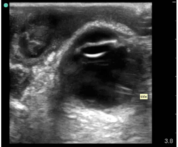
SonoCase: 30 yr old with pelvic pain, LMP 5 weeks ago, stable vitals, mild tenderness in suprapubic area, pelvic exam normal. Your Transabdominal pelvic view on ultrasound shows the below. What do you do next?

Well, we see a full bladder – which is a must if you actually care about your pelvic views – and we see a gestational sac with a double decidua sign – the earliest sign of an intrauterine pregnancy, BUT it is not enough to diagnosis a definitive intrauterine pregnancy – you need a yolk sac within your gestational sac to say that! So, get your endocavitary ultrasound probe and take a look (after emptying the bladder), you may just see the yolk sac!

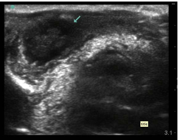
SonoCase: 24 yr old with pelvic pain, LMP 6 weeks ago, stable vitals, mild tenderness in suprapubic area, pelvic exam normal. Your Transabdominal pelvic view on ultrasound shows below. What is the diagnosis? What do you do next?


Well, there is an empty bladder (unfortunately), so the visualization is not its best, but while we slowly fan through the pelvis there is a fluid filled circular cystic-like structure with mild acoustic enhancement (brightness deep to it) which allows you to also see a gestational sac within the uterus. There is an ovarian cyst. Is that what’s causing the pelvic pain? Are you sure it’s not an ectopic? Not yet – get your endocavitary probe now that the bladder is empty and take a look for the yolk sac or fetal pole. If the fetal pole has cardiac activity visualized then we can say it is a LIVE intrauterine pregnancy.

SonoStudy: Prehospital Chest Ultrasound matters!

In a recent study published in Journal of Emergency Medicine, there is a high powered study with quite a few patients on whether prehospital chest ultrasound changes management, destination, or intervention. Once again, a great study highlighting how point of care ultrasound should be used by prehospital providers.

“BACKGROUND:

Due to advancements in technology, the use of a portable ultrasound (US) machine in the out-of-hospital setting is increasingly feasible. It has diagnostic and therapeutic advantages and may improve the management and treatment of patients. It can be used in-flight and can be easily taught to flight clinicians who have little previous experience with this modality.

STUDY OBJECTIVES:

The goal of this study was to evaluate the impact of ultrasound chest examinations on the care of patients treated by a Helicopter Emergency Medical Service (HEMS).

METHODS:

Since 2007, portable US has been used by the HEMS of Nijmegen, The Netherlands. Data on every air medical flight are routinely collected in a database. Every portable US examination of the chest performed between 2007 and 2010 was reviewed for this study. Data on patient characteristics, properties of US examinations, US diagnoses, and impact on medical treatment were collected and analyzed.

RESULTS:

Of a total of 2572 patients, 326 portable US examinations of the chest were performed on 281 (11%) patients. The mean duration of a portable US examination was 2.77 (SD 1.30) min, and the duration decreased over time. After the US examination, the plan for treatment changed in 60 (21%) patients. In 10 patients (4%) the plan to place a chest tube was abandoned. In 10 patients (4%) the initially selected destination for definitive care changed, and it changed to a lower-level hospital more often than to a higher-level one. In 9 patients (3%), cardiopulmonary resuscitation was stopped and in 31 patients there were other changes.

CONCLUSION:

Out-of-hospital US examinations can alter and improve treatment decisions and destinations for definitive care.”

SonoStudy: Flat IVC predictor of poor prognosis in trauma – A-B-C-D-Echo in Trauma!

A recent study on the IVC and trauma and acute surgical patients was done by Ferrada et al, and despite the giggles I get when I read it and how they describe the IVC as “Fat ” or “Flat”, it is an important topic to discuss as it is one of the few articles out there that correlate the iVC to trauma patients and acute surgical patients. First off, it is a retrospective study, which can make it difficult to assess patients with similar factors without other factors coming into play (but when is that NOT the case, honestly?) They did compare the IVC in all patients and studied those patients who seemed sick as well – ICU admission, immediate surgery need, transfusion needed. The power of the study was good but not great – 101 patients studied – varying in type of trauma and surgical need. There was a previous study published in the Journal of Trauma in 2011 that stated CT evidence of flat IVC was an indicator for hypovolemia and  poor prognostic indicator for blunt solid organ injuries – this confirmed a study done in 2010 stating the same thing. Thankfully, ultrasound can get you that information much more immediately than CT!

This month, another study by the same author (Ferrada) in the Journal of trauma and acute care surgery entitled A-B-C-D-Echo (I know, love it!) stated that adding limited transthoracic echo, including the IVC, will benefit trauma patients with results showing “Flat inferior vena cava was associated with an increased incidence of ICU admission (p < 0.0076) and therapeutic operation (p < 0.0001). Of the 148 patients, 27 (18%) had LTTE results indicating euvolemia. The diagnosis in these cases was head injury (n = 14), heart dysfunction (n = 5), spinal shock (n = 4), pulmonary embolism (n = 3), and stroke (n = 1). Of the patients, 121 had LTTE results indicating hypovolemia. Twenty-eight hypovolemic patients had a negative or inconclusive Focused Assessment with Sonography for Trauma examination finding (n = 18 penetrating, n = 10 blunt), with 60% having blood in the abdomen confirmed by surgical exploration or computed tomographic scan. Therapy was modified as a result of LTTE in 41% of cases. Strikingly, in patients older than 65 years, LTTE changed therapy in 96% of cases.”

Below are the Abstracts of the studies highlighted by Ferrada:

“Flat inferior vena cava (IVC) on ultrasound examination has been shown to correlate with hypovolemic status. We hypothesize that a flat IVC on limited echocardiogram (LTTE) performed in the emergency room (ER) correlates with poor prognosis in acutely ill surgical patients. We conducted a retrospective review of all patients undergoing LTTE in the ER from September 2010 until June 2011. IVC diameter was estimated by subxiphoid window. Flat IVC was defined as diameter less than 2 cm. Fat IVC was defined as diameter greater than 2 cm. Need for intensive care unit admission, blood transfusion requirement, mortality, and need for emergent operation between patients with flat versus Fat IVC were compared. One hundred one hypotensive patients had LTTE performed in the ER. Average age was 38 years. Admission diagnosis was blunt trauma (n = 80), penetrating trauma (n = 13), acute care surgery pathology (n = 7), and burn (n = 1). Seventy-four patients had flat IVC on initial LTTE. Compared with those with fat IVC, flat patients were found have higher rates of intensive care unit admission (51.3 vs 14.8%; P = 0.001), blood transfusion requirement (12.2 vs 3.7%), and mortality (13.5 vs 3.7%). This population also underwent emergent surgery on hospital Day 1 more often (16.2 vs 0%; P = 0.033). Initial flat IVC on LTTE is an indicator of hypovolemia and a predictor of poor outcome.”

ABCDEcho:

“BACKGROUND: Limited transthoracic echocardiogram (LTTE) has been introduced as a technique to direct resuscitation in intensive care unit (ICU) patients. Our hypothesis is that LTTE can provide meaningful information to guide therapy for hypotension in the trauma bay.

METHODS: LTTE was performed on hypotensive patients in the trauma bay. Views obtained included parasternal long and short, apical, and subxyphoid. Results were reported regarding contractility (good vs. poor), fluid status (flat inferior vena cava [hypovolemia] vs. fat inferior vena cava [euvolemia]), and pericardial effusion (present vs. absent). Need for surgery, ICU admission, Focused Assessment with Sonography for Trauma examination results, and change in therapy as a consequence of LTTE findings were examined. Data were collected prospectively to evaluate the utility of this test.

RESULTS: A total of 148 LTTEs were performed in consecutive patients from January to December 2011. Mean age was 46 years. Admission diagnosis was 80% blunt trauma, 16% penetrating trauma, and 4% burn. Subxyphoid window was obtained in all patients. Parasternal and apical windows were obtained in 96.5% and 11%, respectively. Flat inferior vena cava was associated with an increased incidence of ICU admission (p < 0.0076) and therapeutic operation (p < 0.0001). Of the 148 patients, 27 (18%) had LTTE results indicating euvolemia. The diagnosis in these cases was head injury (n = 14), heart dysfunction (n = 5), spinal shock (n = 4), pulmonary embolism (n = 3), and stroke (n = 1). Of the patients, 121 had LTTE results indicating hypovolemia. Twenty-eight hypovolemic patients had a negative or inconclusive Focused Assessment with Sonography for Trauma examination finding (n = 18 penetrating, n = 10 blunt), with 60% having blood in the abdomen confirmed by surgical exploration or computed tomographic scan. Therapy was modified as a result of LTTE in 41% of cases. Strikingly, in patients older than 65 years, LTTE changed therapy in 96% of cases.

CONCLUSION: LTTE is a useful tool to guide therapy in hypotensive patients in the trauma bay.”

SonoStudy: Serial compression DVT ultrasound with doppler of iliacs can rule out DVT in pregnancy

In a recent Medscape article, a study is highlighted regarding serial DVT ultrasound (including doppler of iliacs) can rule out DVT in pregnancy. How about that?! This is a follow up to that last post regarding “best test” for first diagnosis of suspected DVT. The article is stated below:

“Conducting serial compression ultrasonography with Doppler imaging of the iliac vein over the course of 7 days can rule out deep-vein thrombosis (DVT) in symptomatic pregnant women, according to a prospective cohort study.

Wee-Shian Chan, MD, from the Department of Medicine, BC Women’s Hospital and Health Centre, Vancouver, British Columbia, Canada, and colleagues report their findings in an article published online January 14 in the Canadian Medical Association Journal.

Increasingly, healthcare providers suspecting DVT turn to serial venous compression ultrasonography to diagnose the blood clots; if negative, Doppler imaging is added to overcome ultrasound’s lack of sensitivity in diagnosing isolated iliac DVTs in pregnant women for whom pelvic veins cannot be compressed. The diagnostic approach is noninvasive, widely available, and does not expose the fetus to ionizing radiation, and thus has become the standard of practice for diagnosing DVT in pregnant women. Because no study has validated this approach, Dr. Chan and colleagues sought to evaluate the diagnostic accuracy of using serial compression ultrasonography and Doppler imaging of the iliac veins among symptomatic pregnant women during a 7-day period.

They focused on pregnant women consecutively referred for investigation of DVT from August 2002 to September 2010 at 4 Canadian centers and enrolled 221 women who had had a suspected first episode of DVT. Exclusion criteria included receipt of anticoagulant therapy for more than 48 hours before presentation, suspicion of pulmonary embolism, concomitant cardiac or pulmonary disease, and inability or lack of willingness to return for follow-up.

Trained technicians performed ultrasound examination of the symptomatic leg by compression of the proximal veins (ie, along the length of the femoral vein from the inguinal canal and the popliteal veins to the level of the calf trifurcation with Doppler studies of the iliac vein). If the first test results were negative for DVT, blood thinners were withheld and testing was repeated after 2 to 4 days and again after 6 to 8 days. Blood thinners were prescribed if results were positive. The research team excluded DVT based on full compressibility of the femoral and popliteal veins and normal Doppler imaging of the iliac veins.

“The overall prevalence of deep vein thrombosis in our study cohort was 7.7% (95% [confidence interval,] 4.9% – 12.0%),” Dr. Chan and colleagues write. “The iliac or femoral veins, or both, were involved in 65% (11/17) of cases. Of these, 2 (12%) cases were isolated to the iliac vein, and 4 (24%) were isolated to the femoral vein.”

Some 94% of DVT were detected by the initial compression ultrasonography with Doppler imaging. The serial testing that followed did not detect any new thromboses, the research team writes. “Our strategy of serial compression ultrasonography combined with Doppler imaging of the iliac veins appears to reliably exclude clinically important deep vein thrombosis,” the authors write.

Added Value Unclear

Additional studies are needed to determine what is better for pregnant women: ultrasonic testing with Doppler imaging or single whole-leg ultrasound, “the only other diagnostic strategy that has been formally validated for DVT suspicion in pregnancy,” Grégoire Le Gal, MD, from the University of Brest, France, told Medscape Medical News. Dr. Le Gal and colleagues found that conducting a single ultrasound test may rule out diagnosis of DVT in pregnant and postpartum women.

“In nonpregnant patients, the 2 strategies have been compared and showed similar safety. The use of single whole-leg ultrasound is convenient, because patients don’t need to come back for serial testing. But, on the other hand, it is more time-consuming and it leads to a positive diagnosis of DVT in a higher proportion of patients, mainly because all distal DVT are detected — and therefore treated — with anticoagulants, when this approach is used,” Dr. Le Gal says. Using serial compression ultrasonic testing, “only the distal DVT that extend proximally are detected on serial tests and treated.”

Dr. Le Gal also noted that in Dr. Chan’s study, a Doppler interrogation was performed in all patients. “Physicians willing to implement this diagnostic strategy in their clinical practice need to take this into account. Unfortunately, the yield of this test was not provided in the manuscript. Therefore, it is impossible to know the exact added value of this test as compared with a serial compression ultrasonography without Doppler,” Dr. Le Gal told Medscape Medical News.

Because it is unethical to use leg venography with fluoroscopy or computed tomographic angiography in pregnant women to confirm DVT diagnoses, Dr. Chan and colleagues assumed that all diagnosed abnormalities were DVT. Another study limitation was that the current study showed a lower prevalence of DVT compared with 2 previous studies involving pregnant women; this could exaggerate the negative predictive value. In addition, the study investigators were thrombosis specialists based in secondary and tertiary referral centers, which raises the question of generalizability; because patients were recruited from a range of settings, however, the researchers argue that the study’s results are generalizable to most community-based hospitals.

“Our study highlights the importance of iliac vein visualization in symptomatic pregnant women,” the authors conclude. They add that the research also “shows that serial compression ultrasonographic studies with iliac vein visualization performed over a 7-day period can reliably exclude deep vein thrombosis in symptomatic pregnant women and that it is likely safe to withhold anticoagulation in women with negative imaging results.”

CMAJ. Published online January 14, 2013. Full text

SonoStudy: The “best” test for evaluating for first suspected episode of DVT

A recent highlight from Mescape discusses a study published in Annals of Internal Medicine by Linkins et al. about how the D-Dimer may or may not help in DVT evaluation, and how ultrasound relates to the diagnosis of DVT in varying groups based on pre-test probability. Interesting read, and definitely something to make me go “hmmmm….”. The study concludes that in a certain group, a selective d-dimer testing paradigm can be of utility. The Mescape are states:

“It is best to base ᴅ-dimer testing on a patient’s clinical pretest probability (C-PTP) for deep vein thrombosis (DVT), rather than testing all patients who present with symptoms of first DVT episode. This strategy can exclude DVT in more patients without increasing missed diagnoses, according to a randomized, multicenter, controlled trial in 1723 patients at 5 medical centers in Canada.

Lori-Ann Linkins, MD, an assistant professor in the Division of Hematology and Thromboembolism, Department of Medicine, McMaster University in Hamilton, Ontario, Canada, and colleagues published their findings in the January 15 issue of the Annals of Internal Medicine.

ᴅ-dimer testing is sensitive but not specific for identifying DVT. Selectively testing ᴅ-dimer levels lowered the proportion of patients who needed ultrasonography and decreased the percentage of patients who required ᴅ-dimer testing by 21.8% (95% confidence interval [CI], 19.1% – 24.8%).

“In this trial comparing uniform with selective ᴅ-dimer testing in patients with suspected first DVT, a selective strategy — which used a higher ᴅ-dimer threshold to exclude first acute DVT in outpatients with low C-PTP and omitted ᴅ-dimer testing in outpatients with high C-PTP and all inpatients — was as safe as and more efficient than the uniform testing strategy, which used the same threshold to exclude DVT in all patients,” the authors write.

Patients were randomly assigned to the selective testing (n = 860) or uniform testing (n = 863) groups on presentation for suspected first DVT episode. Of the study participants, 1542 (89%) were outpatients and 181 (11%) were inpatients.

All patients in the uniform testing group underwent ᴅ-dimer testing. Levels less than 0.5 μg/mL were considered negative, and levels of 0.5 μg/mL or higher were considered positive. For patients with positive results, ultrasonography of the proximal veins in the symptomatic legs was conducted; patients with normal ultrasonogram and high C-PTP had ultrasonography repeated on the same legs 6 to 8 days later.

Patients in the selective testing group only underwent ᴅ-dimer testing if they were outpatients and had low or moderate C-PTP. Outpatients with high C-PTP and all inpatients underwent ultrasonography only. ᴅ-dimer levels in the low C-PTP group were considered negative if they were below 1.0 μg/mL and positive if they were 1.0 μg/mL or above.

For patients in the moderate C-PTP group, ᴅ-dimer levels were considered negative if they were below 0.5 μg/mL and positive if they were 0.5 μg/mL or above. Patients with positive results had ultrasonography, and patients with normal ultrasonogram and moderate or high C-PTP had ultrasonography repeated 6 to 8 days later.

Of the patients in the uniform testing group, 859 (99.5%) had ᴅ-dimer testing, 505 (58.5%) had initial ultrasonography, and 334 (38.7%) had ultrasonography repeated after 6 to 8 days.

Positive ᴅ-dimer results were found in 506 patients (418 outpatients and 88 inpatients), and negative results were found in 353 patients (351 outpatients and 2 inpatients). Four patients had no test. DVT was diagnosed by initial ultrasonography in 56 ᴅ-dimer-positive patients (11.1% of the 506 ᴅ-dimer-positive patients and 6.5% of 863 patients in the uniform testing group). None of the 81 patients with low C-PTP and a ᴅ-dimer level between 0.5 and 1.0 µg/mL had DVT on ultrasonography.

“You’re Not Missing Cases”

Venous thromboembolism (VTE) was diagnosed during follow-up in 4 patients: 0.8% (95% CI, 0.2% – 2.0%) of the 506 ᴅ-dimer-positive patients with normal initial ultrasonogram and 0.5% (95% CI, 0.1% – 1.3%) of the 798 patients without DVT on initial testing who were still in the study at 3-month follow-up. No outpatients with low C-PTP and ᴅ-dimer levels between 0.5 and 1.0 μg/mL were diagnosed with VTE during follow-up. No VTE was diagnosed during follow-up in any ᴅ-dimer-negative patient (0.0%; 95% CI, 0.0% – 1.1%).

ᴅ-dimer testing was done in 668 of the 860 patients (77.7%), initial ultrasonography was done in 438 patients (50.9%), and ultrasound was repeated after 6 to 8 days in 383 patients (44.5%). ᴅ-dimer results were negative in 288 (80%) and positive in 72 (20%) of the 360 outpatients with low C-PTP.

None of the 288 ᴅ-dimer-negative patients (200 with ᴅ-dimer level < 0.5 μg/mL and 88 with ᴅ-dimer level 0.5 – 1.0 μg/mL) experienced VTE during follow-up (95% CI, 0.0% – 1.3%).

Of the 72 ᴅ-dimer-positive patients, 8 (11%) had DVT diagnosed by ultrasonography during initial testing. No patient with a normal ultrasonogram experienced VTE during follow-up (95% CI, 0.0% – 5.1%).

A total of 132 (43%) of the outpatients with moderate C-PTP were ᴅ-dimer-negative and 176 (57%) were ᴅ-dimer-positive. Two patients had no ᴅ-dimer testing. One ᴅ-dimer-negative patient experienced VTE during follow-up (0.8%; 95% CI, 0.0% – 4.3%), and 5 were lost to follow-up. DVT was diagnosed by ultrasonography during initial testing in 23 (13%) of the 176 ᴅ-dimer-positive patients. One of the 153 ᴅ-dimer-positive patients with normal ultrasonogram developed VTE during follow-up (0.6%; 95% CI, 0.0% – 3.2%); 4 patients were lost to follow-up.

DVT was diagnosed during initial testing in 20 (10.5%) of the 100 outpatients with high C-PTP and the 90 inpatients. VTE was identified during follow-up in 2 patients with normal ultrasonograms (1.1%; 95% CI, 0.1% – 3.8%). One patient was lost to follow-up.

DVT was diagnosed during initial testing in 51 (5.9%) of the selective testing patients. VTE developed during follow-up in 4 (0.5%; 95% CI, 0.1% – 1.3%) of the patients who had no DVT diagnosed during initial testing.

During follow-up, the difference between the groups in the number of VTE events was 0.0 percentage points (95% CI, −0.8 to 0.8 percentage points) in patients not diagnosed with DVT during initial testing and −0.3 percentage points (95% CI, −1.8 to 0.8 percentage points) in favor of selective testing in the outpatient or low C-PTP subgroup.

The difference between the groups in the proportion of those undergoing testing was −21.8 percentage points (95% CI, −24.8 to −19.1 percentage points) for ᴅ-dimer testing and −7.6 percentage points (95% CI, −12.2 to −2.9 percentage points) for ultrasonography, both in favor of selective testing.

The proportion of patients in the outpatient and low C-PTP subgroup who had ultrasonography was 20.0% in the selective testing group and 41.0% in the uniform testing group (difference, −21.0 percentage points in favor of selective testing; 95% CI, −27.6 to −14.2 percentage points).

Daniel J. Giaccio, MD, vice chair of medicine at Lutheran Medical Center in Brooklyn, New York, commented on the study in a telephone interview with Medscape Medical News. Selective testing enabled the researchers to avoid unnecessary ultrasounds, he noted. “In this day and age of cost-effectiveness, and especially with the incentives…for doctors to practice more cost-effective care, [it’s good] to know that you’re not missing cases — you’re actually picking up more,” Dr. Giaccio explained.”

SonoStudy: >29,000 patients: Utility of cardiac portion of FAST scan: should we be doing it?

Should we keep doing the echo with the FAST scan? What does it truly add? Ill never forget the story I heard about a 35 yr old male blunt trauma victim after single vehicle motor vehicle accident who lost his pulse en route.  The echo part of the FAST scan showed tamponade in the first 5 minutes of evaluation and ACLS/ATLS management. He survived due to early pick up) and walked out of the hospital. Or, the penetrating epigastric stab wound victim who was tachycardic and hypotensive with no tamponade or pericardial effusion seen on FAST (helping us rule out tamponade as the cause of shock). But, when looking at the studies….a recent one from JEM states:

“Background

Focused assessment with sonography in trauma (FAST) is widely used and endorsed by guidelines, but little evidence exists regarding the utility of the cardiac portion in blunt trauma. The traditional FAST includes the routine performance of cardiac sonography, regardless of risk for hemopericardium.

Study Objectives

Our goal was to estimate the prevalence of hemopericardium due to blunt trauma and determine the sensitivity of certain variables for the presence of blunt hemopericardium.

Methods

We performed a retrospective chart review of two institutional databases at a large urban Level I trauma center to determine the prevalence of blunt hemopericardium and cardiac rupture and incidental or insignificant effusions. We evaluated the sensitivity of major mechanism of injury, hypotension, and emergent intubation for blunt hemopericardium and cardiac rupture.

Results

Eighteen patients had hemopericardium and cardiac rupture (14 and 4, respectively) out of 29,236 blunt trauma patients in the Trauma Registry over an 8.5-year period. The prevalence was 0.06% (95% confidence interval [CI] 0.04–0.09%). The prevalence of incidental or insignificant effusions was 0.13% (95% CI 0.09–0.18%). One case of blunt hemopericardium was identified in the emergency ultrasound database out of 777 cardiac ultrasounds over a 3-year period. No patient with blunt hemopericardium or cardiac rupture presented without a major mechanism of injury, hypotension, or emergent intubation.

Conclusion

Blunt hemopericardium is rare. High-acuity variables may help guide the selective use of echocardiography in blunt trauma.”

So, I would ask: is it worth the 20 seconds it takes to look at the heart to pick up those patients who had a positive scan? And, is it worth the 20 seconds it takes to look at the heart to rule it out? – i say – yes. But I get it – it may be negative A LOT of the time.

SonoPearls&Politics: ACEP2012, AAMC2012, AIUM-US First- future of bedside ultrasound

2012 was an amazing year for bedside ultrasound. There were more conferences that included bedside ultrasound in their pre-conferences festivities, but also there were more discussions on what was next for bedside ultrasound, while SUSME and AIUM announced 2013 as the Year of Ultrasound (YOU) – highlighted by AIUM Ultrasound First group, the Life in the Fast Lane bloggers, the Ultrasound Podcast folks, and, of course, little ole’ me on SonoSpot while highlighting the ACEP US Section and the immense amount of social media interest/bloggers/tweets on the topic of bedside ultrasound. There are two conferences I went to, each with it’s own powerful voice with regard to education, medicine, and ultrasound. The excitement I felt was truly unprecedented – I was giddy, I was hopping around, I was all smiles.

The American College of Emergency Physicians (ACEP) meeting had more ultrasound lectures and workshops than ever before with a turnout at the ACEP US Section that was more than any other (although I dont have the exact numbers, the ballroom it was held in was huge, and those who came late had to stand because all the seats were filled). ACEP was amazing. period. From the great lectures/workshops (even the on-site resuscitation of an emergency physician who went into cardiac arrest in the lobby of the convention center (revived by fellow emergency physicians through use of the handy-dandy convention center defibrillator to then have his heart checked for cardiac activity by Dr. Chris Fox with the ultrasound machine he was using during his workshop, which was happening right next to that location) and the Aurora Mass Casualty Response Video, (also seen here), which was one of the most moving videos I’ve seen about emergency response, teamwork, and humanity (I’ve said this many times, but Ill say it again – I LOVE my job – but even better than that, I love those who I do my job with – side-by-side – and what a privilege to be able to feel that way) to everything inbetween and afterwards, ACEP was once again a success.

The Association of American Medical Colleges (AAMC) meeting in San Francisco was equally amazing, particularly with regard to the future of medical education, discussing the concept of the flipped classroom, and the time given to discussing the incorporation of bedside ultrasound into medical education for medical schools – with the first ultrasound workshop being held in its history lead by the “God’s of Ultrasound in MedEd” (that’s my term of choice)  -Drs. Richard Hoppmann (Univ South Carolina), Chris Fox (UC Irvine), and Michael Blaivas (all of whom will be at the World Congress: Ultrasound in Med ED)…. with help from ultrasound educators from Wayne State, Ohio State, and Stanford (yup, little ‘ole me again and my star medical student models). There was even a separate day at Stanford where a 60 minute slot was given to discussing The Stanford 25 (by none other than Stanford’s Dr. Abrahim Verghese himself) and one of it’s aspects, Bedside Ultrasound (by one of our ultrasound team members, Dr. John Kugler, an internal medicine doctor who is starting to incorporate ultrasound into internal medicine residency education – yup, it’s spreading!! – and it’s about time!). No tweets on this conference, but the above should be stated anyway.

The Ultrasound First conference went on with tweets happening every hour! I was unable to attend this one, but so happy that my twitter friends did. It is obvious that 2013 truly is the year of ultrasound. Spreading to medical education, being a multi-disciplinary educational and practical tool, and having a united voice on its value were all discussed – in addition to some pearls on the hot topics including pelvic ultrasound and MSK ultrasound, as well as how ultrasound is becoming an acceptable tool for renal colic and breast masses.

Since I learn from all my Twitter friends, I figured the best way to share is to take out the middle person (yes, Im talking about me). That way you could get it from their own words: Here are only a few posts from #ACEP12  and #US1st that made me go “Hmmmm….” – with a little commentary every now again from me, because I just can’t NOT give my opinion – I know that’s shocking to those of you who know me. Heehee. My tweets are in here as well.

ACEP:

From @USEDCDN : Emergency US management course  “From Blaivas: Starting to see 1st lawsuits for lack of US use in vascular access” – This definitely sparked my attention – lawsuits for LACK of US use?? Wow, well the standard of care is changing, and if a proven tool to minimize complications is right next to you and you dont use it and that complication occurs… there’s a legal risk. Learn it, use it, love it and maximize patient safety.

Also from  “@USEDCDN: EUS MC Resnick: Emergency US is not an extension of physical exam. Big difference. It answers clinical questions.” Ok, this needs mention, but I already posted a rant about this – of course – so will not bother you with another rant… not right now, at least.

From @jeremyfaust  – “Weingart: 4. When is CPR futile? End tidal < 10 after 10 min. Confirm with US #acep12.” Enough said – and honestly, anything that Scott Weingart says, i will believe. period.

“Rice #ACEP12 echo in cardiac arrest- can see if cause PE/tamponade, or if standstill or beating heart. Look or you may waste time/resources” and “echo and IVC in critical patients: LV fxn, IVC collapse, RV size, contractility- will differentiate PE, CHF, hypovolemia, hypervolemia” – you never know what you may find, and what may be an intervention that you didnt think of until you saw your ultrasound (ie. tpa in a dilated RV).

@bedsidesono: lung #ultrasound talk from ACEP athttps://vimeo.com/51212231  brush up on A-Lines, B-Lines, lung sliding and more…#FOAMed” – what a giver he is! Stone is one to listen to, hear his opinion, and read his immense amount of publications.

Point of care US dominating new speakers forum so far at #ACEP12! Msk, soft tiss, pleural…”

Congrats @GeriaSonoMD on being new Chair of #ACEP12 US section mtg. Our fellow, Viveta Lobo said you talked her into EM. Awesome. So thx!

Raj Geria – new #ACEP12 US section Chair! Highest priority: pathway to US fellowship accreditation : to ensure safety and quality

Find @SAEMAEUS on twitter and follow to see what’s new with the ACademy

Nova panebianco at #ACEP12 taking about SAEM Academy of Emerg US and SonoGames – also subcommittees they are involved in..

A great resource from the new academy of emerg US : http://SAEM.org/academy-emergency-ultrasound-resources …

Resa Lewis #ACEP12 US section mtg- talking ACGME US milestones and how ACEP can help in achieving them for all residencies

Blaivas #ACEP12 – TEE will show potential causes of hypotension & shock ..Valvular dz..And can be electrically linked :pace & defibrillate. No need to interrupt chest compressions for TTE as can see what’s happening with heart from across rm c TEE. Can use TEE when bad view on TTE or unable to do TTE due to habitus, lung dz, chest compressions. TEE can assess quality of chest compressions too. TTE can tell you if there is standstill, clot in RA, dilated RV, tamponade, and to see a beating heart. TTE (echo) better than checking for pulses for need for chest compressions. AAMC mtg in SF! Spreading to med schools!. WINFOCUS and AIUM EM and crit care goals… Going global!

http://Sonocloud.org  and http://sonoguide.com  : 2 great online resources for images and education! – plus the test: http://emsono.com
@sinaiemus: Rob Blankenship at Ultrasound section meeting: over 56,000 ACEP US tests completed at http://www.emsono.com/acep/ACEP_EUS_Exam.html … #ACEP12
Congrats Vicki Noble and @ultrasoundpod for your well deserved award for your contribution to emerg ultrasound!!
Ultrasound First Forum:
  1. Jason T Nomura MD @Takeokun “To engage the patient groups you need people who are interested in patient advocacy not just the disease state. 
  2. View image on Twitter
  3.  Jason T Nomura MD @Takeokun “Lev demonstrating high res eval of ankle tendons with dynamic scanning for function, something that can’t be done with MRI 
  4. Jason T Nomura MD @Takeokun “Nazarian MRI does not have the resolution to evaluate the fibrillar pattern of the Achilles compared to US. 
  5. Jason T Nomura MD @Takeokun “Hoppmann- if education and integration of US starts in medical school it can change the paradigm. 
  6.  Jason T Nomura MD @Takeokun “Hoppmann has graduated several classes of medical students who had US integrated into their med school curriculum. 
  7. Jason T Nomura MD @Takeokun “Moreau most common imaging modalities for Team USA is X-ray and US, very little CT use.
  8. Mike Stone @bedsidesono “Levon Nazarian at  speaking on MSK imaging. It’s not just more convenient than MRI – higher res, no contraindications, pt’s prefer it”
  9.  Jason T Nomura MD @Takeokun “Nazarian US for sports med is portable to get the technology to the field and locker room, MRI not portable. 
  10. Jason T Nomura MD @Takeokun “Pellikka 2011 joint guidelines from ACC,ASE, ACCP and others about the appropriate times to use echo in the assessment of pts. 
  11.  Jason T Nomura MD @Takeokun “Moore bringing up the ASE and CV Anes guidelines for US guided vasc access. Advocates real time US guidance 
  12. Mike Stone @bedsidesono “Leslie Scoutt from Yale – ACR appropriateness criteria for recurrent renal colic – US & Noncon CT equal ratings 
  13.  Jason T Nomura MD @Takeokun “Scoutt 50% of pts with renal colic will likely have another episode.  that rad exp can build up.”
  14.  Jason T Nomura MD @Takeokun “Scoutt noncon CT is the “gold standard” for renal colic imaging in the US currently.  but there is the rad “risk.
  15. Joshua Copel @jacopel “Lynn Fordham (Pedi Rads) US optimal for pyloric stenosis now. No more need for upper GI or other radiation. 
  16. Jason T Nomura MD @Takeokun “Fordham N/V can be pyloric stenosis, malro, intussusception, and gastroenteritis. US for dx.
  17.  Jason T Nomura MD @Takeokun “IOTA group from Europe with close to 2,000 pts showed very good discrimination of malignant vs benign ovarian mass on US. 
  18. Jason T Nomura MD @Takeokun “IUD placement or misplacement easy to note on US; can present for DUB and pain. t
  19. Jason T Nomura MD @Takeokun “Advances to 3D US allows volumetric imaging that could only be done previously with CT or MRI. But US spares the radiation of CT 
  20. Joshua Copel @jacopel “ Dr. Beryl Benacerraf making case for US over CT, MR in female pelvic imaging at forum. pic.twitter.com/vCQYvpi8 View image on Twitter
  21. Jason T Nomura MD @Takeokun “ is not only about when &where US can be used but education for practitioners and patients per @AIUMPresAlfred
  22. Jason T Nomura MD @Takeokun “@AIUM_Ultrasound represents 9,200 members from 36 specialties with a focus on advancing US use 
  23.  Joshua Copel @jacopel “ opening of US First forum now at Marriott NYC. Over 100 attending from medical profs, industry, payors. Very exciting & energetic”
  24.  Jason T Nomura MD @TakeokunIt does seem to be a who’s who of US at the reception.

SonoStudy: A time-series analysis: central IV rate after US-guided peripheral IV program

A recent study in the Annals of EM by Shokoohi et al did a time series analysis of the rate of central line requirements after an US-guided peripheral IV program was implemented – 80% reduction! you read that right…. read on for the abstract:

“Study objective
We examine the central venous catheter placement rate during the implementation of an ultrasound-guided peripheral intravenous access program.

Methods
We conducted a time-series analysis of the monthly central venous catheter rate among adult emergency department (ED) patients in an academic urban ED between 2006 and 2011. During this period, emergency medicine residents and ED technicians were trained in ultrasound-guided peripheral intravenous access. We calculated the monthly central venous catheter placement rate overall and compared the central venous catheter reduction rate associated with the ultrasound-guided peripheral intravenous access program between noncritically ill patients and patients admitted to critical care. Patients receiving central venous catheters were classified as noncritically ill if admitted to telemetry or medical/surgical floor or discharged home from the ED.

Results
During the study period, the ED treated a total of 401,532 patients, of whom 1,583 (0.39%) received a central venous catheter. The central venous catheter rate decreased by 80% between 2006 (0.81%) and 2011 (0.16%). The decrease in the rate was significantly greater among noncritically ill patients (mean for telemetry patients 4.4% per month [95% confidence interval {CI} 3.6% to 5.1%], floor patients 4.8% [95% CI 4.2% to 5.3%], and discharged patients 7.6% [95% CI 6.2% to 9.1%]) than critically ill patients (0.9%; 95% CI 0.6% to 1.2%). The proportion of central venous catheters that were placed in critically ill patients increased from 34% in 2006 to 81% in 2011 because fewer central venous catheterizations were performed in noncritically ill patients.

Conclusion
The ultrasound-guided peripheral intravenous access program was associated with reductions in central venous catheter placement, particularly in noncritically ill patients. Further research is needed to determine the extent to which such access can replace central venous catheter placement in ED patients with difficult vascular access.”

Great videos for Peripheral venous access:

And, to watch an installment of the UltrasoundPodcast of their IV course, watch here.

SonoCase: Pulling Sensation in the Eye – by EPMonthly: Drs. Teresa Wu and Brady Pregerson

In the most recent issue of EP Monthly, Drs. Teresa Wu and Brady Pregerson, give another enlightening and humorous story of the average every-day emergency patient after first describing how they explained to their EM intern the role of emergency physicians (which I couldnt agree more with!): “As an EM physician, you are a healer, an educator, a detective, a diagnostician, and a master strategist all rolled into one.”

The case:

“41-year-old male who presents to the ED with concerns that his left eye is progressively getting more swollen. He’s had some increasing eye pain and purulent drainage over the past six days. At first he thought that he was just having really bad seasonal allergies, but today, he started feeling a “pulling sensation” on the medial aspect of his left eye. He denies any headache, diplopia, sinus pain, rhinorrhea, nausea, vomiting, or recent trauma. He does note a subjective fever at home, and his temperature is 38.2°C in the ED. His vital signs otherwise demonstrate tachycardia to 123 bpm, but a normal blood pressure, oxygen saturation, and respiratory rate. Your intern has asked the nurse to obtain a visual acuity on the patient and he is systematically going through his ocular exam when you walk by the room to check on him. He comes out of the room to give you an update on what he’s discovered so far. The patient has tenderness to palpation over his left medial orbit and possible entrapment on ocular exam. He has no additional pain with extraocular movement and no diplopia, but has so much periorbital edema that it wasn’t possible to get a consistent Tonopen measurement. There doesn’t appear to be any fluorescein uptake on the slit lamp exam, and other than conjunctival injection and the lid swelling, the patient has a normal ocular exam.

It is now about 4:30 pm and you know that in 30 minutes, all consultants turn into pumpkins and their pagers magically stop working. As you are about to ask your intern what he wants to do next, the medical student pulls up the ultrasound machine that the intern asked her to wheel over and hands it to him. He takes the linear array transducer and performs an ocular ultrasound at the bedside. He saves the following images: what do you see?”

Screen shot 2013-01-07 at 1.57.18 PM

Screen shot 2013-01-07 at 1.57.22 PM

To find out…..and read up on what it was, what happened, and the pearls of the exam, go here.

SonoSocial: Social Media in EM Ultrasound Education – check out what’s out there!

Happy Monday everyone! Im sure you love mondays as much as we do, so i thought I would post something that would be a bit of positivity and highlight a group that I believe in: ACEP Ultrasound Section. 2013 is the YEAR OF ULTRASOUND – and for good reason – there are only a few tools that give us such immediate information that can save a life. The ACEP US Section is the go-to site for everything you want to know about starting an US program, credentialing in ultrasound, the policies and politics, and is the home of SonoGuide – an amazing educational resource for bedside ultrasound, and the EMSONO: Ultrasound Test. It is also where we add our entries for their newsletter that goes over tips and tricks, cases, and all things ultrasound in the news. We recently wrote an article for the ACEP Ultrasound Section Newsletter – which is available for all members of the ACEP US Section – and I highly recommend becoming a member – it’s totally worth it. It is my go-to guide for all things bedside ultrasound. I thought to entice you, i would include our latest entry below – with a few additions in the end as it was written a few months ago and there have been more educators/bloggers since it’s publication: For a set of links to online education in bedside ultrasound, go here.

Social Media in EM Ultrasound Education by Drs. Viveta Lobo, Laleh Gharahbaghian

Stanford University Medical Center

In a realm where the access to free education, is becoming the mantra and expectation across learners, the emergence of social media is fast gaining popularity. A quick Google search, defines social media as, “a media for social interaction, using highly accessible and scalable publishing techniques.” As a physician, educator, and learner in an academic institution, this modality of education promises to bestow unending means. Being able to have a presence 24/7 and allow access to learning at the convenience of learners is almost too good to be true. It is no wonder why higher education across the globe, and now even primary education has adopted social media as the forefront modality to reach and connect with learners.

The medical field is no exception. As we begin to recruit the most “tech savvy” group of students in our medical schools and residencies, the expectation to continue learning “on the cloud” is ongoing. Tweeting anatomy pearls, sharing Dropboxfiles on physiology notes, and joining Google hangouts for journal clubs – is quickly becoming the theme for most educational sites. And why shouldn’t it be? Being able to remotely participate in learning and connect with peers anywhere in the world, from your home, office, or emergency department, can only lead to extraordinary things.

Emergency medicine has rightly developed a reputation for being progressive and innovative. Emergency ultrasound lends itself to be the perfect subset to thrive with social media, and it has taken off! Being able to upload ultrasound clips online and teach anatomy, highlight pathology, and reward “great saves” for not just your own institution, but the entire ultrasound community goes way beyond the classroom. Websites, Blogs, Podcasts, Facebook group pages – whatever your fancy, its there – and it’s free. Our goal, has always been to try, learn, fail, succeed – but at the end, share. There is no better way to share than by using social media. Being able to connect all across the globe with other EM US enthusiasts, interact and discuss various topics and strategies has only helped us all do better, within our sub specialty and beyond. So don’t be skeptical, and give it a try! We’ve listed some of our favorite FREE picks, in no particular order, to help you get started…. There are plenty more out there….Until next time – see you on the cloud!

Websites for Online Education/Didactics in Bedside Ultrasound:

Sonoguide: Ultrasound Guide for Emergency Physicians – offers a description of each bedside US application, ultrasound images and videos – all to train and review US utility for aiding diagnosis, management and treatment by Dr, Beatrice Hoffman

SonoWorld – a comprehensive site that includes hundreds of lectures, cases, videos, and articles that includes basic bedside ultrasound applications, but goes beyond that to satisfy the needs of all US enthusiasts.

Emergency Ultrasonography – a comprehensive website geared to emergency physicians that contains many lectures, cases, and educational content that can be used by residencies for the didactic portion of ultrasound education by Dr, Geoff Hayden.

SAEM Ultrasound Narrated Lectures – just as it states, a library of lectures on bedside ultrasound with a link to a free pdf of ultrasound guides on that same site.

Sinai EM Ultrasound – a complete site of free online lectures, cases, research articles, and news on bedside ultrasound topics by Dr. Bret Nelson.

Society of US in Medical Education – learning modules and curriculum databases with a long lecture list on all basic bedside ultrasound topics that allows the learner to control the pace of the lecture.

Vanderbilt’s EM Ultrasound – a collection of brief lectures on each bedside ultrasound topic in a well-organized way with quizzes that can be taken to test your knowledge by Dr. Jim Fiechtl

ICU Sonography lectures for Echo/IVC and more by: Beth Israel, Stanford, and Yale

Online Textbooks on Bedside Ultrasound:

European Federation of Societies for Ultrasound in Medicine and Biology (EFSUMB)– a free and comprehensive European full textbook found online for free on each ultrasound application

Partners In Health Textbook for Ultrasound in Resource Limited Areas – by Drs. Sachita Shah, Dan Price, Gene Bukhman, Sachin Shah, Emily Wroe

iBook for the iPAD – a new textbook brought from the Ultrasound Podcast guys and written by many US educators sure to bring a new concept to the world of online education – The iBook and all it’s glory: http://www.youtube.com/watch?v=mB7_omzi8Cc

Podcasts on Bedside Ultrasound:

Ultrasound Podcast – one of the best, and funniest ways of teaching ultrasound to all learners. This podcast discusses all ultrasound applications, keeps it easy, and makes it engaging by Drs. Mike Mallin and Matt Dawson, with Stone’s side excerpts from Dr. Mike Stone.

UC Irvine’s iTunes lecture series – an excellent and comprehensive lecture podcast library of all bedside ultrasound applications by Dr. Chris Fox.

Soundbytes – a collection of free lectures for all bedside ultrasound applications in a concise and free format by Dr. Phil Perera.

Blogs on Bedside Ultrasound:

SonoSpot: Topics in Bedside Ultrasound – a blog that discusses tutorials, cases, tips/tricks, research, links/sites, and people in the ultrasound community by Dr. Laleh Gharahbaghian (yes, shameless plug here).

Bedside Ultrasound – Ultrasound teaching updates with tips/tricks and brief video lectures of the hot topics in bedside ultrasound and interviews with US enthusiasts by Dr. Mike Stone.

Highland Hospital ED Ultrasound – a collection of blog posts that focus on procedural US guided applications in addition to the basic US application by Dr. Arun Nagdev and Andrew Herring

Ultrasound videos & clips:

Sonocloud – the ultimate library of ultrasound clips – an amazing free image upload and download site where you can find any ultrasound clip you need to review, place in your lecture, or use to learn and teach others by Drs Mike Mallin, Matt Dawson, and Mike Stone.

Vimeo: US in Emergency Medicine and Critical Care group – a large collection of ultrasound clips for your viewing pleasure of pathologies, normal anatomy, and more by the HQMedEd (high quality medical education) team at Hennepin County Medical Center

There’s an App for That! – free Smartphone apps for bedside ultrasound

1-Minute Ultrasound – by the Ultrasound Podcast guys for your iPhone, with 1 minute reviews of all applications in an easy to learn way.

SonoAccess – a list of lectures and images in a smartphone app by SonoSite

Twitter accounts to follow for bedside ultrasound teaching topics: 123SonographyAIUM_Ultrasound, AllUltrasound, Bedsidesono, BMUS_Ultrasound, ECCUltrasound, EchoCardiac, EDUltrasound, EDultrasoundQA, EM Res, GeriaSonoMD, SAEMAEUS, Sinai EM Ultrasound, SonoSpot, SUSME, UltrasoundFirst, UltrasoundMeme, UltrasoundPod, Ultrasound Training, Ultrasound Quarterly, USEDCDN

Other SonoBelievers who contribute to ultrasound education through their blogs/tweets:

Academic Life in EMLife in the Fast Lane, EMCRIT, CriticalCareNow, HQMedEdSonoCaveSonoIntensivist, WesternSono, TeresaWuMD, ChrisPartyka, AriKestler, CurroMiralles, Radiopaedia, EMCurrents, Jason T Nomura, Squartadoc, AndyNeill, EMManchester, EMBasic, AllUltrasound, BroomeDocs, M-Lin, EMEducation, EMChatter, Precordialthump, I_C_N, MDAware, EMIMDoc, jvrbntz, SandnSurf, IMEducator, keeweedoc, LWestafer, ERCast, DocWagz

A new, up and coming site for free, fun-incentivized global medical education through cases, questions, images (including ultrasound): Global Medical Education Project

I know…. a big family!… and I guarantee you Im missing a few, but there will be even more in 2013: The Year of Ultrasound.