SonoLectures: Free lecture on Ultrasound in the Critically Ill -by Dr. Cliff Rice (& other free lectures)

Got an email from ACEP and thought it was too good not to share: Hear Dr. Cliff Rice, an ultrasound extraordinaire and emergency physician speak about bedside ultrasound and its use in critical care medicine. At the end of this post are even more lectures that are free. As you will hear, he states “Think about how you would use it in some of our sickest patients that come to the emergency department….. where the differential diagnosis is quite broad, and the treatment for shock might be detrimental if we are wrong.”

Screen Shot 2013-03-04 at 4.12.42 PM

As ACEP states in the email: “Practicing emergency physicians need to be able to utilize ultrasound effectively in the evaluation of the critically ill patient. In this free audio recording from the 2012 ACEP Scientific Assembly, Dr. Rice highlights the use of ultrasound to perform a FAST scan, to dynamically monitor and measure the IVC in the setting of hypovolemic shock, and to detect pericardial effusion and perform ultrasound guided pericardiocentesis [in 45 minutes]. This [lecture] explains where you should start scanning, narrows your differential and guides your resuscitation.”

Other free lectures for your viewing/hearing pleasure on bedside ultrasound:

Dr. Chris Fox’s comprehensive emergency ultrasound lectures in iTunes

Dr. Phil Perera comprehensive emergency ultrasound lectures on Sound-Bytes

AIUM UltrasoundFirst lecture series on various ultrasound topics

UltrasoundPodcast with a variety of lectures on bedside ultrasound

UltrasoundVIllage website on a variety of ultrasound topics

Vanderbilt’s excellent lectures library on bedside ultrasound

SonoWorkshop: Pearls (and more!) from the Stanford CME Ultrasound Course #FOAMed

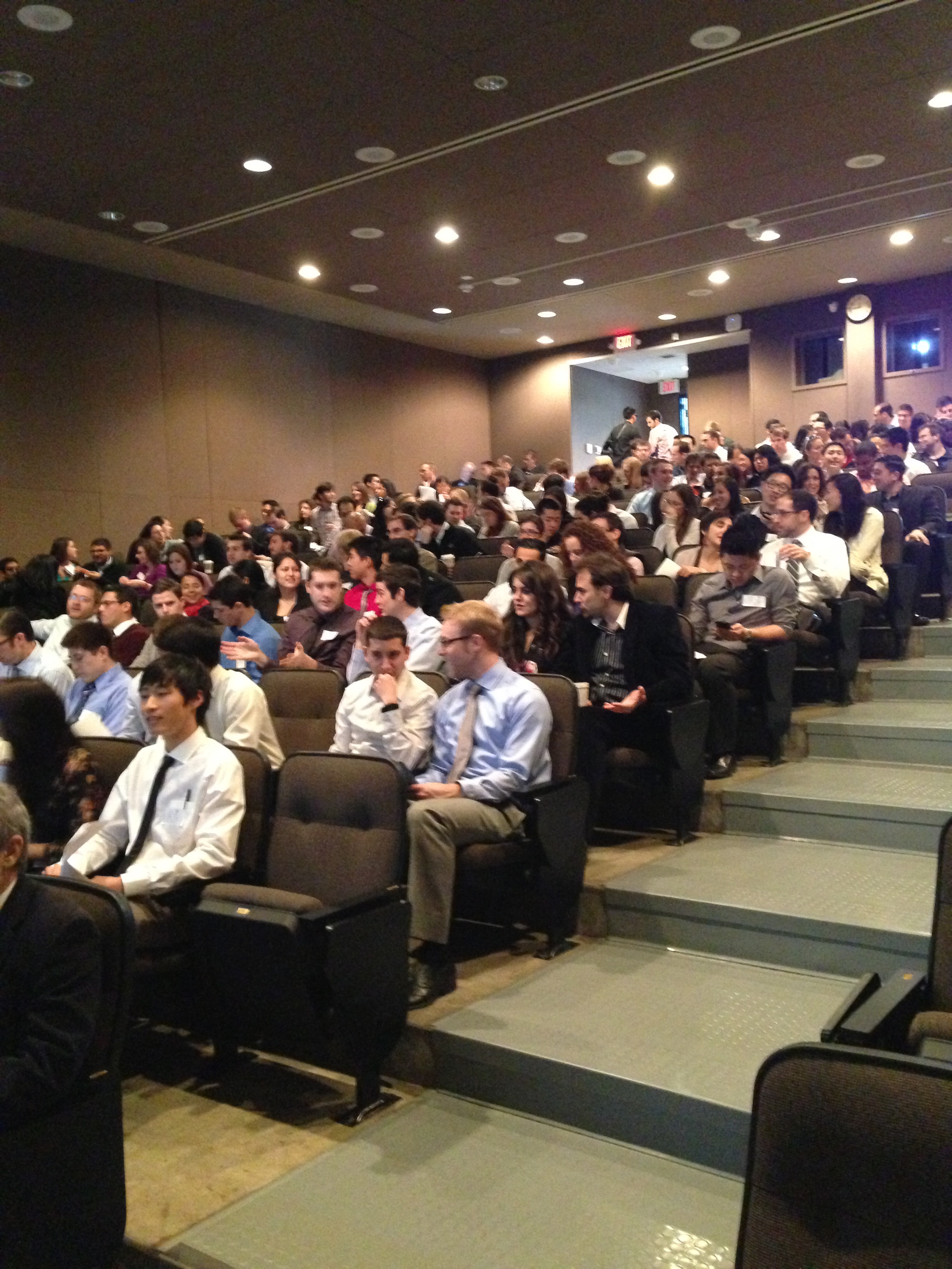
Once again, our Stanford Ultrasound Workshop was a huge success. Why? Our instructors were phenomenal and from different specialties! Our participants were faculty from emergency medicine, internal medicine, critical care, surgery, and pediatrics! The ultrasound tips and tricks just kept on coming from our lecturers  – and, everyone laughed at our jokes, which always makes things great. As always, I like to provide those tips and tricks to all of you (and maybe even some of the jokes), so that you can feel like you were there too!

IMG_1087

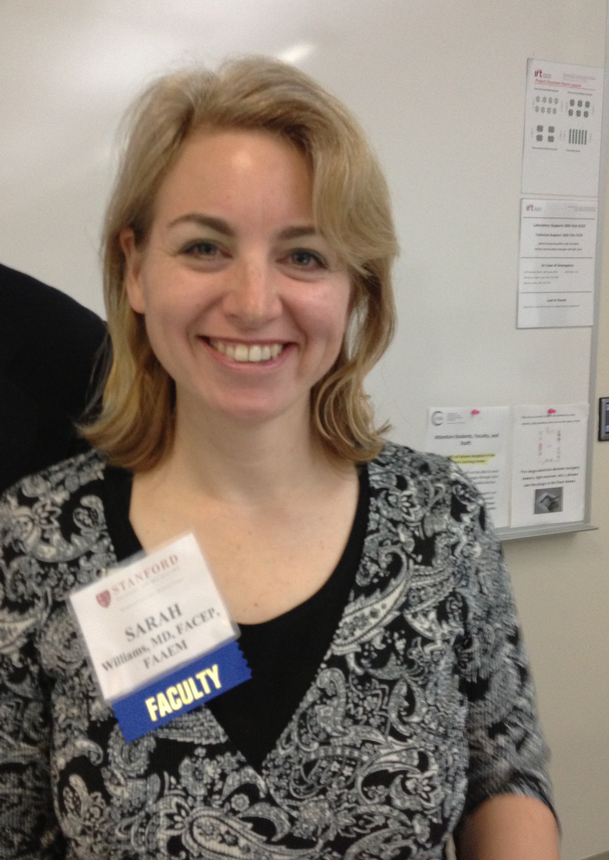
Dr. Sarah Williams – First, the coordinator for the Stanford CME workshop welcomes everyone with a Star Wars phrase “Learning you are….May the force be with you, young padawans!” – always goes to a great start. She is also the creator of the Stanford Ultrasound Program and current Associate Residency Director (and the person who was kind enough to put up with my quirks and jokes to hire me as a fellow years ago). Her pearls on the EFAST: detects >600 cc (intraperitoneal) fluid, look around inferior pole in RUQ and subdiaphragm area of LUQ (free fluid develops first there!), it’s not good for pelvic fx/injuries (pelvic bleeding into pelvic cavity, and retroperitoneal, bowel gas obstructs view, bladder may be empty limiting visualization), it’s not done fast- FAST is part of RUSH, but dont rush the FAST. Look for your kidney, then look above it, around it and below it (thoracic fluid, morison’s pouch, paracolic gutter). FAST LUQ: higher, spleen smaller, stomach big -place knuckles on gurney, oblique probe in plane to ribs, free fluid can be between diaphragm & spleen.  #ultrasound detects 15-20cc fluid in thoracic cavity, better than chest Xray. Have patient take deep breath to lower diaphragm. The longer the patient is supine (or trendelenberg) the better, so if you have a walk-in trauma, perform serial FAST scan. SX view: the liver is the heart’s protector, be sure to see it in view- it allows you to see the 4chambers. gas is heart’s enemy – if gas gets in the way, you cannot see the liver: slide probe laterally to patient’s right, get that liver in your view. For pneumothorax eval – use linear probe, find your ribs, ID pleur liine, decr gain (brightness) to see sliding better. Start high in midclav line, indicator to head – – then travel thru mult rib spaces to estimate size.

IMG_1089

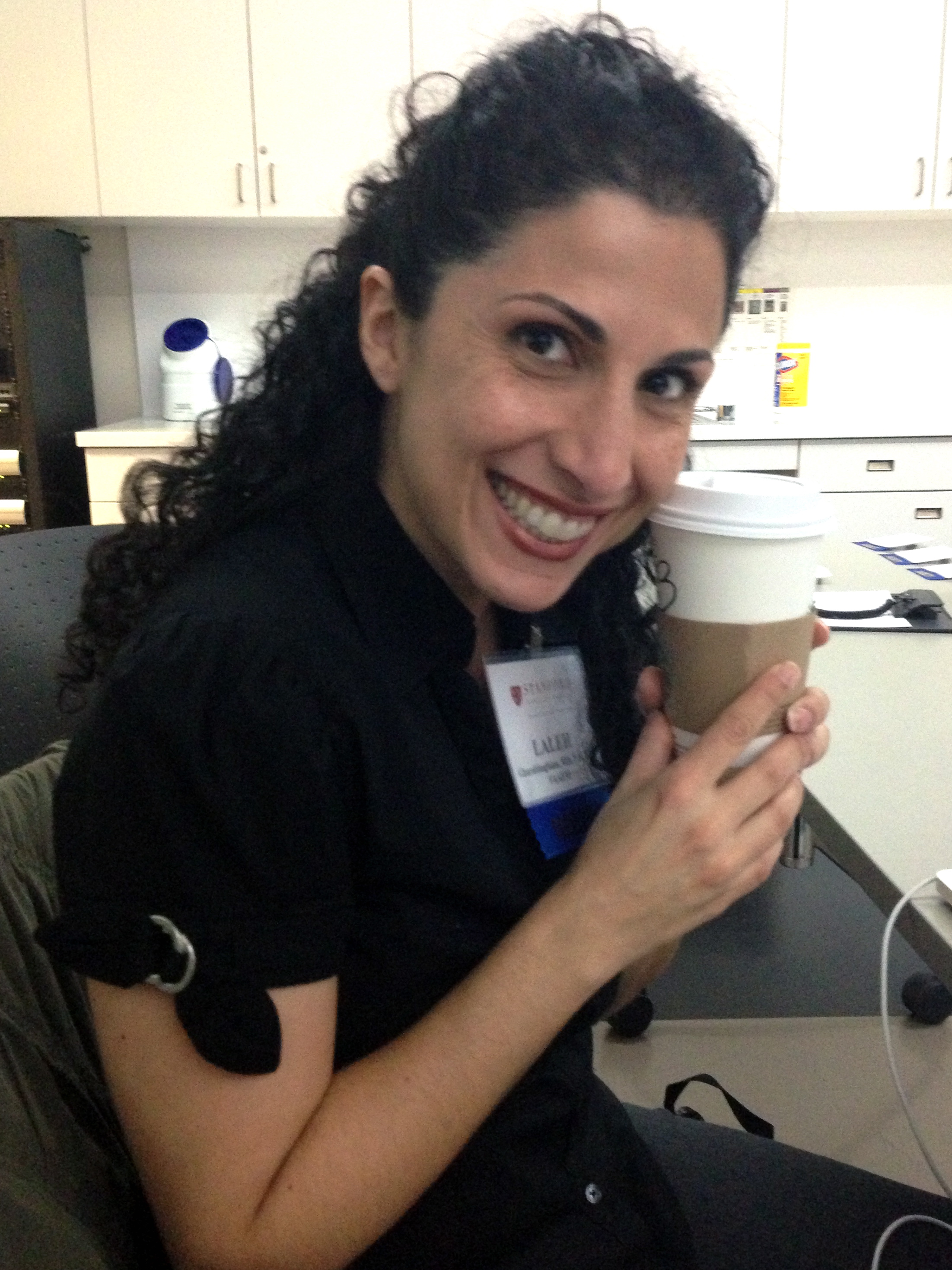
Dr. Laleh Gharahbaghian (since i cannot speak about myself, I let someone else write this part and promised I wouldn’t change anything – let us pray…): “With her usual stylish self, walking all around the workshop, giving hi-fives to everyone in her path, her dance moves came in handy as she spoke (can you point her out in this video from the mid 1990s of her past job?)  – She is the current Director of the Stanford Ultrasound Program and Fellowship her pearls can be found below: Her pearls of Aorta US: use large footprint probe, if get gas, press down, takes time – as if you were reducing a hernia; start in the subxiphoid region, travel down thru to iliacs. Most AAA are infrarenal and may seem normal in size at sx and get large once you travel down. Doesnt evaluate for rupture – most AAA leak/rupture retroperitoneal – not detected by US (your FAST is neg) – correlate clinically to your patient symptoms and vital signs. Her pearls on Renal US: main indication: hydronpehrosis, but pay attention to everything (outside to inside); eval both kidneys AND bladder- without bladder, you wont know if the bilateral hydro may just be that they have to pee. If empty bladder, and bilateral hydro, then possible mass (if not chronic). If patient is >50yo with flank pain, dont forget to eval the aorta as well. Start outside to inside for pathology – free fluid around kidney, cyst from kidney, mass on kidney, stone within kidney, hydronephrosis. Her pearls on Gallbladder US: start in the subxiphoid region, indicator to patient’s right, use liver as window, fan thru it medial to lateral to find GB. Then, fan/eval in transverse & longitudinal planes. Fanning thru the GB is key- there’ll be sections where it looks normal, then you fan & a stone comes into view! See if the stone is mobile by turning patient and re-scanning to see if moved. Think of the number 4 (or multiples of it) with measurements: width 4cm, length 8-10cm, anterior GB wall <4mm, CBD 4mm at 40yrs old (adding 1mm for every decade beyond).

IMG_1088

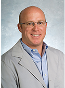
Dr. Phil Perera – our newest addition to the Stanford US team serving as the Director of US Research and the Associate Program Director gave quite an engaging talk on Echo and the RUSH protocol, putting it to action! As is highlighted by his Soundbytes website that is a free source of lectures for your viewing pleasure, he would keep asking the audience whether they would involve their consultants, if they would “write home to mom about this?!” The funny part is that one of the audience members responded with “I wouldn’t have to, she would call me before I get a chance.” Another participant concluded the point by saying, “You must work in a profitable community hospital.” Ah – gotta love the sarcasm! Phil continued his talk discussing when you should act fast by going through RUSH cases, giving props to others who also study and educate on resuscitation ultrasound. His pearls on EchoPSL view is the favorite – lets you evaluate right ventricle size, left ventricle size and contractility, pericardial effusion, pleural effusion and mitral valve regurge; Echo should be done with IVC when thinking about fluid resuscitation – if hyperdynamic -can tolerate fluid; if hypocontractile, not so much; Echo can eval aorta too! PSL view visualizes ascending aorta and descending aorta; AP4 view shows descending aorta – look for aneurysm/flap. Intraperitoneal fluid and pleural effusion can be mistaken for pericardial effusion – know where your pericardium is! Pleual effusion in PSL view travels behind descending aorta; pericardial effusion travels in front of descending aorta. AP4 great for comparing RV and LV chamber size, contractility of RV and LV. To get the P4 view, slide lateral after parasternal views until get to apex, angle to body center. His pearls on RUSH: Case that inspired him: 67yo acute SOB, in shock h/o COPD/CHF/HTN, CXR neg, ultrasound showing the cause to not be sepsis, but cardiogenic shock. RUSH provides the answer to : sepsis? cardiogenic? hypovolemic? hypervolemic? tamponade? PE? trauma? tension ptx? AAA? First & most important is the cardiac echo: the PUMP, that’s why it’s first – lots of info from a single cardiac view (PSL). For semi-quantitative contractility eval: fractional shortening & EPSS are measured – PSL must be at approp long section.  tamponade on #ultrasound – RV collapse during when it should fill (diastole)-also can see RA scalloping -do pericardiocentesis. pericardiocentesis: US studies show having pt in left lateral decubitus position & an apical view better for removing pericardial effusion than traditional SX technique. IVC – can use M mode to measure in both transv and long view 2cm from RA – can use your internal jugular as an alternate. Lung ultrasound – B Lines – think of fluids and your resuscitation when evaluating etiology of shock: FALLS protocol by Lichtenstein. Although rare, if your EKG has STEMI, do an ECHO – make sure its not a dissection before you start heparin!

IMG_1091 and Screen Shot 2013-03-01 at 1.41.03 PM

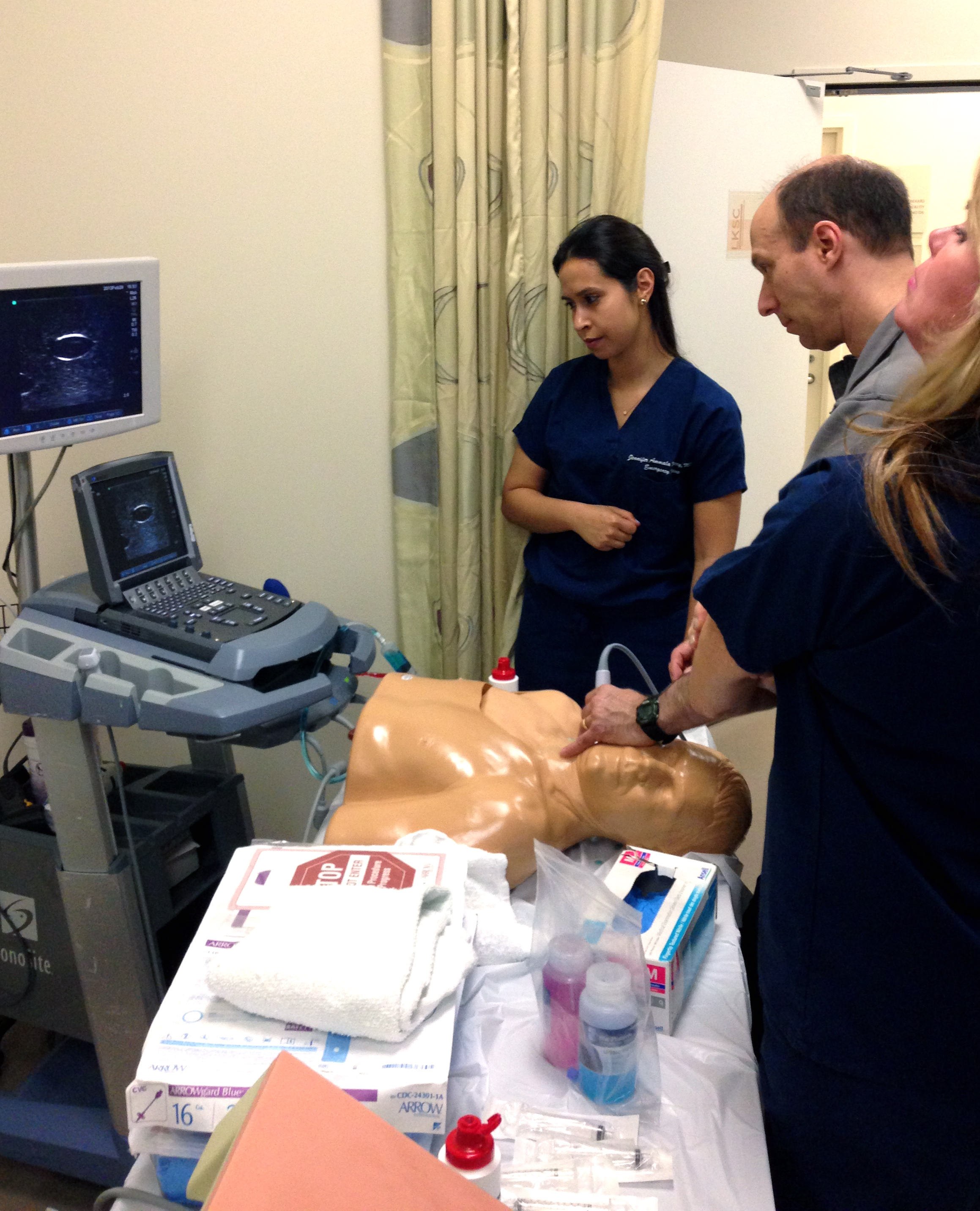
Dr. Zoe Howard (our Director of Medical Student and Resident US Education) and Dr. John Kugler (coordinator for the internal medicine US elective and global health US instructor) spoke about the many awesome ways ultrasound can help with procedural guidance. The dynamic duo had awesome videos to assist in their lecture and went through the many procedures that can be done with US guidance. Their pearls on Procedural US: On central line access – first look for the vein before you prep the area; it’s possible that the vein you want (or the location of the vein you want) is not the best vein for the procedure. Your indicator should be to your left, the screen dot should be on your left, that way left means LEFT when you’re guiding your needle tip to the vein. On lumbar puncture – do it when you can’t feel the landmarks, when you only have one attempt, when you’ve already had one unsuccessful attempt. Use the ALiEM trick with a paperclip for drawing the straight line. On thoracentesis and paracentesis – make sure you view the area where there is at least 2cm of fluid between the probe and the lung/bowel to avoid lung/bowel puncture – it may not always be where you think. On pericardiocentesis – look for where the fluid is most, patient to left lateral decubitus position, and you’ll find that SX is not the best anymore. On nerve blockslearn it, do it, and teach it! Your patients deserve it! Use the in-plane approach to visualize your entire needle, and use the dental syringe holder to have control over your syringe.

IMG_1093 Screen Shot 2013-03-01 at 2.05.40 PM Screen Shot 2013-03-01 at 2.03.42 PM Screen Shot 2013-03-01 at 2.04.57 PM Screen Shot 2013-03-01 at 2.06.47 PM Screen Shot 2013-03-01 at 2.59.35 PM Screen Shot 2013-03-01 at 3.07.32 PM

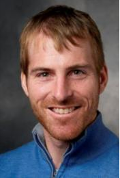
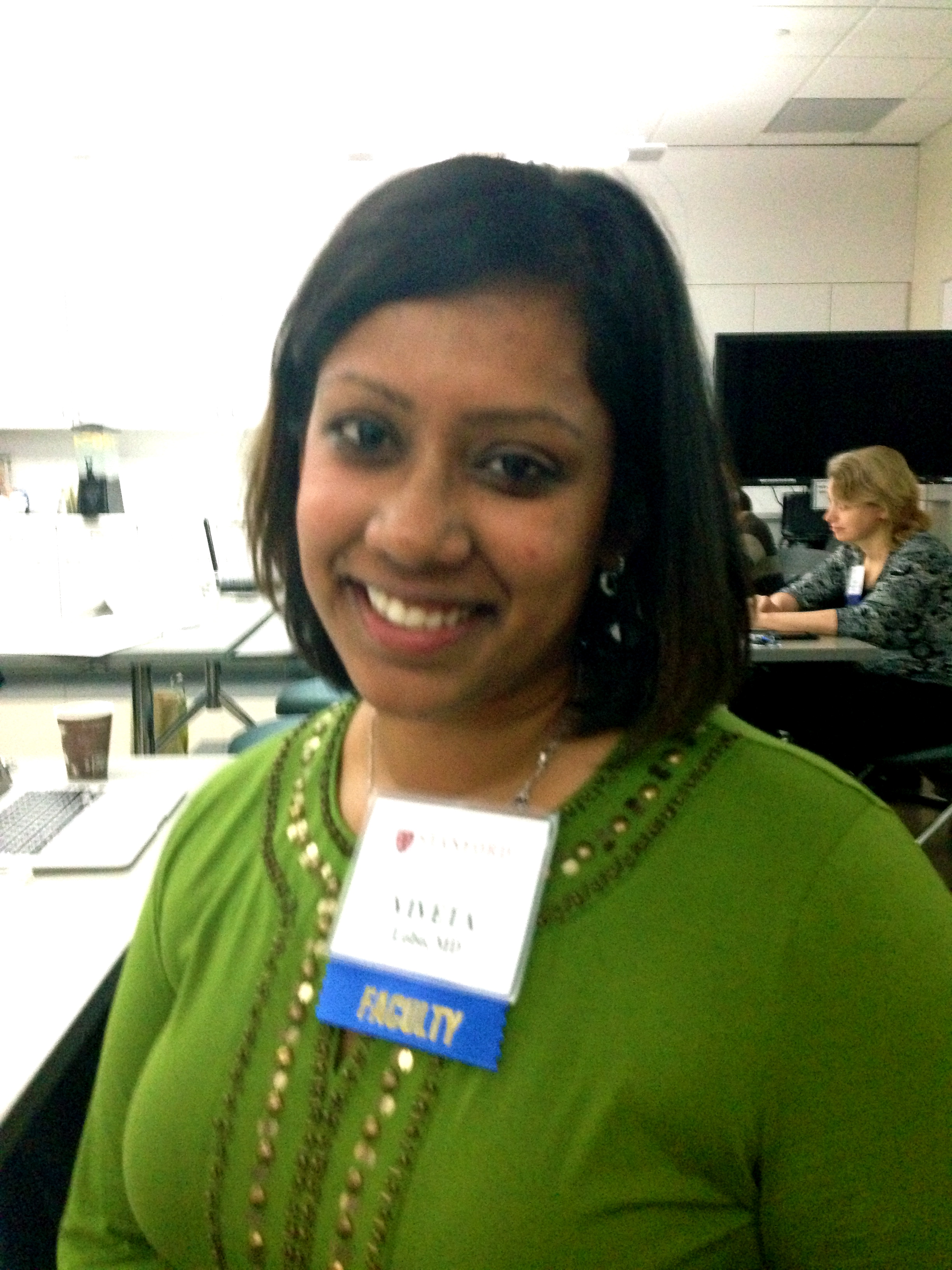
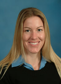
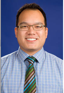
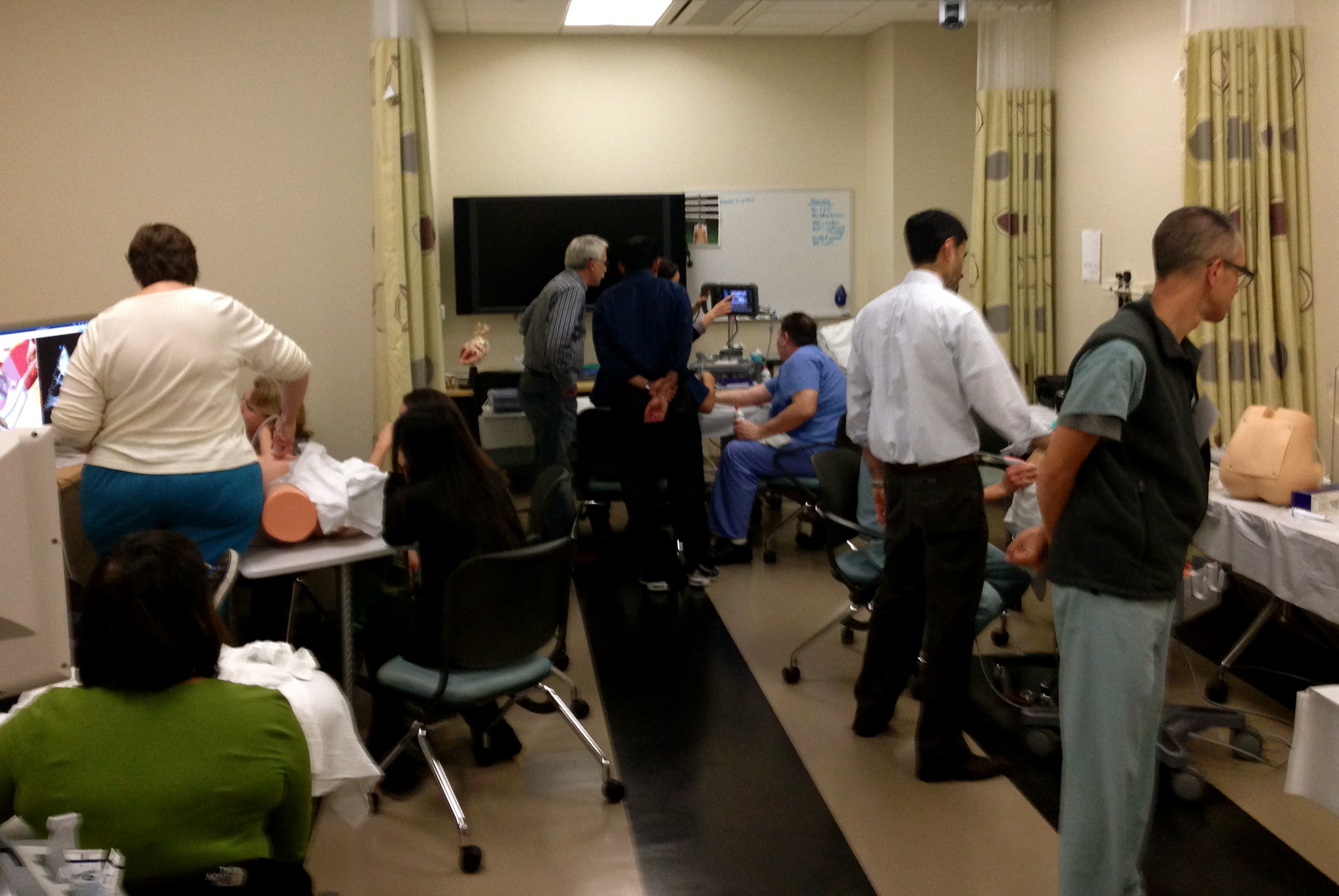
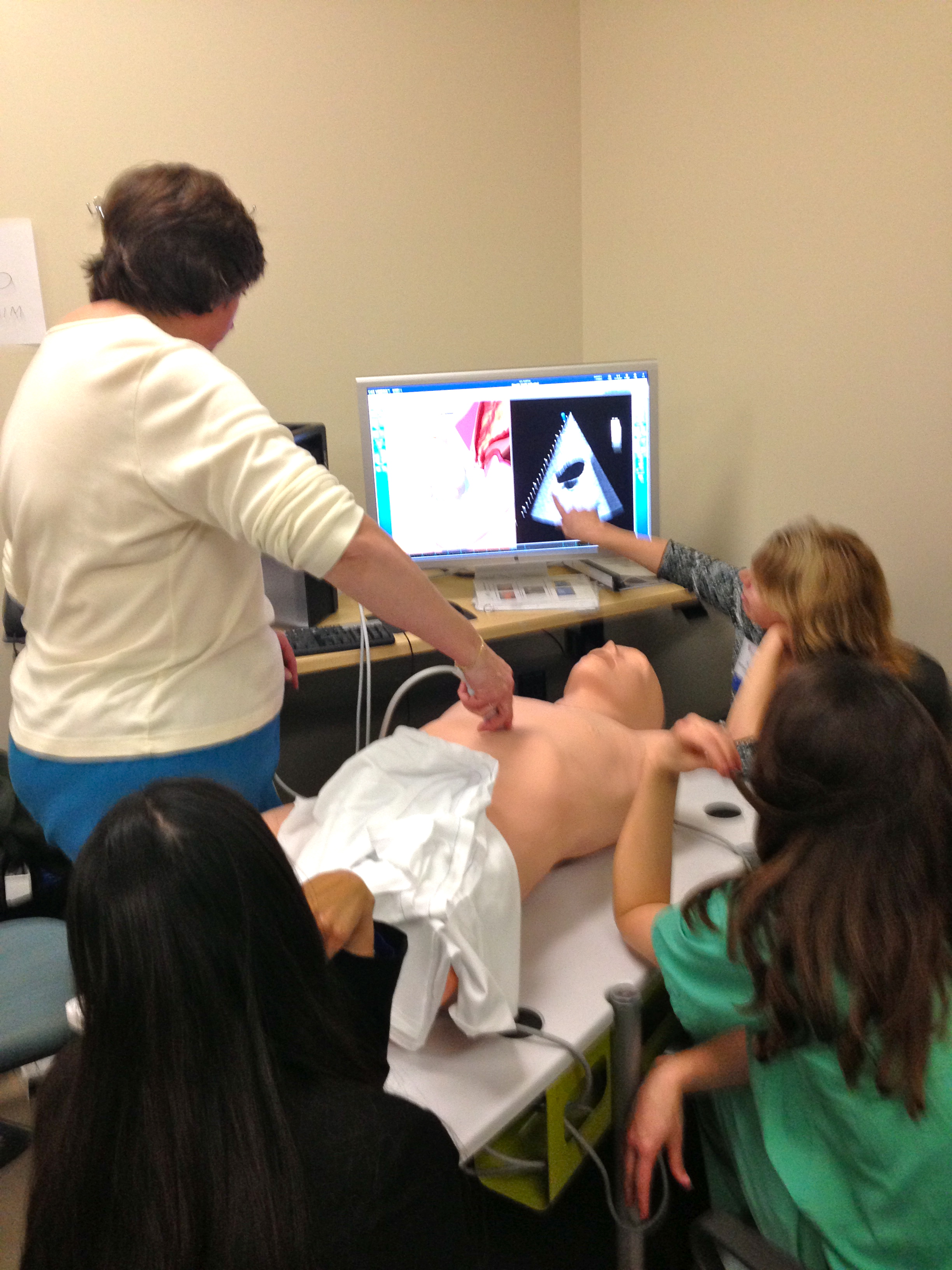
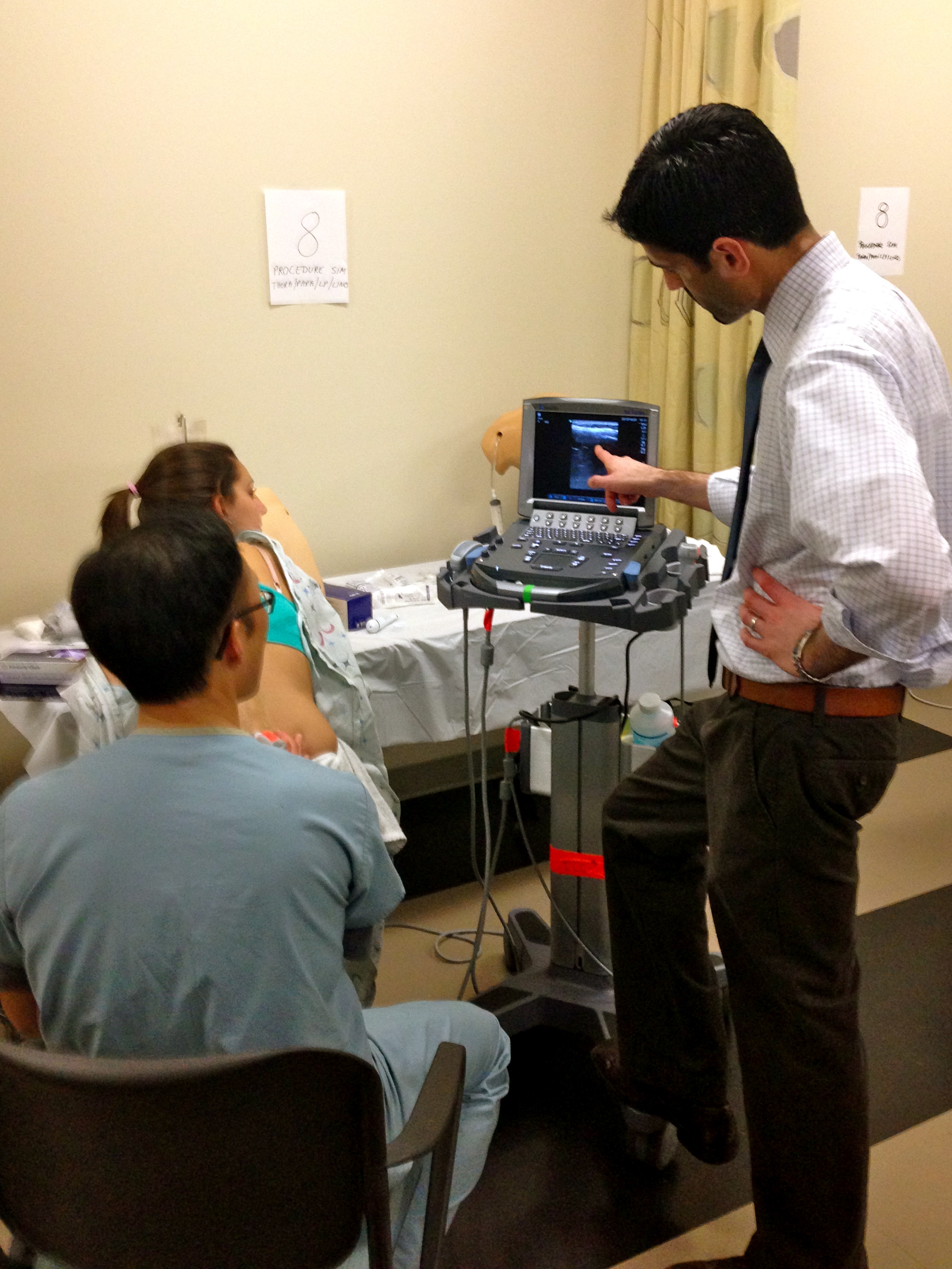
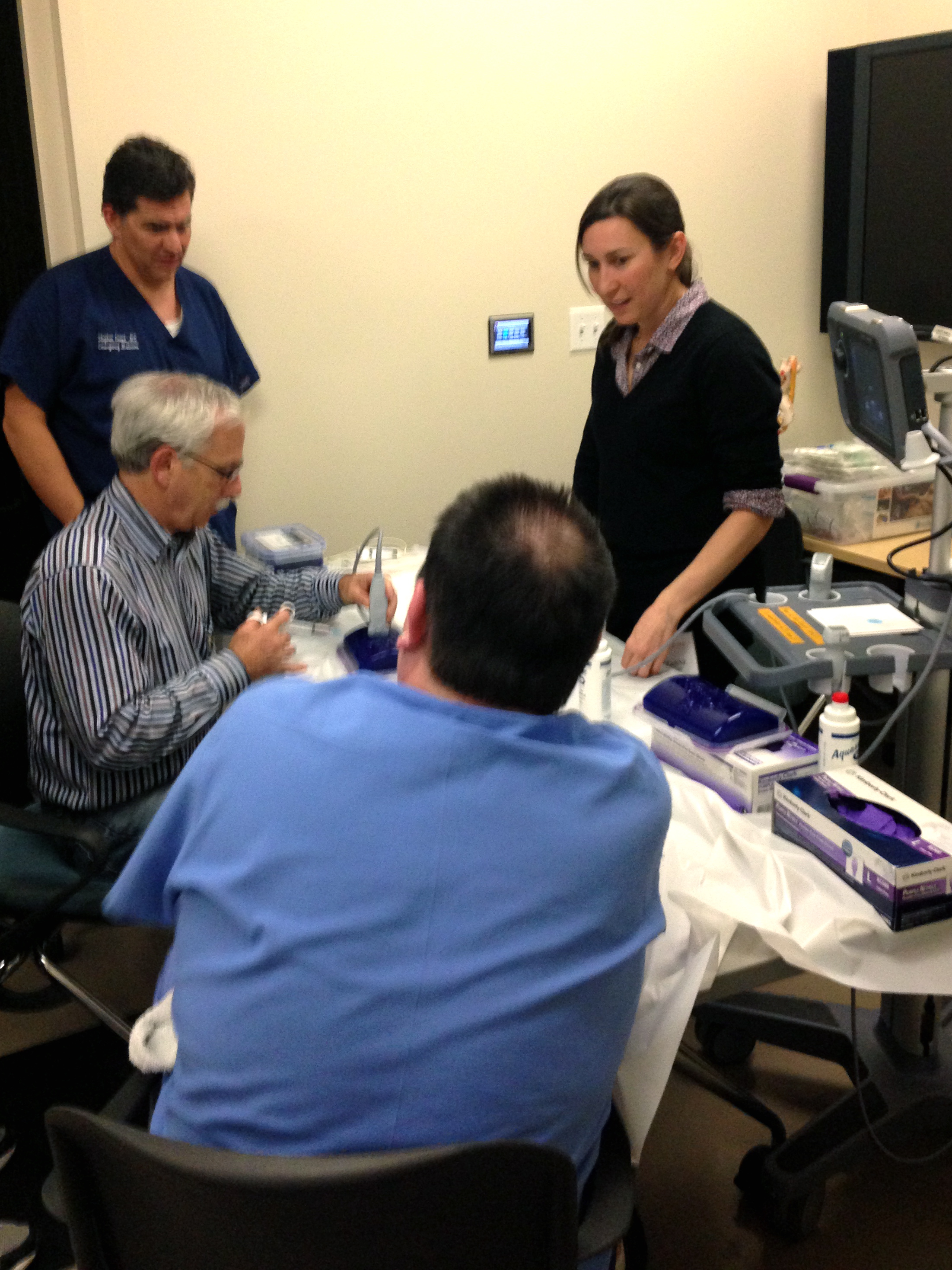
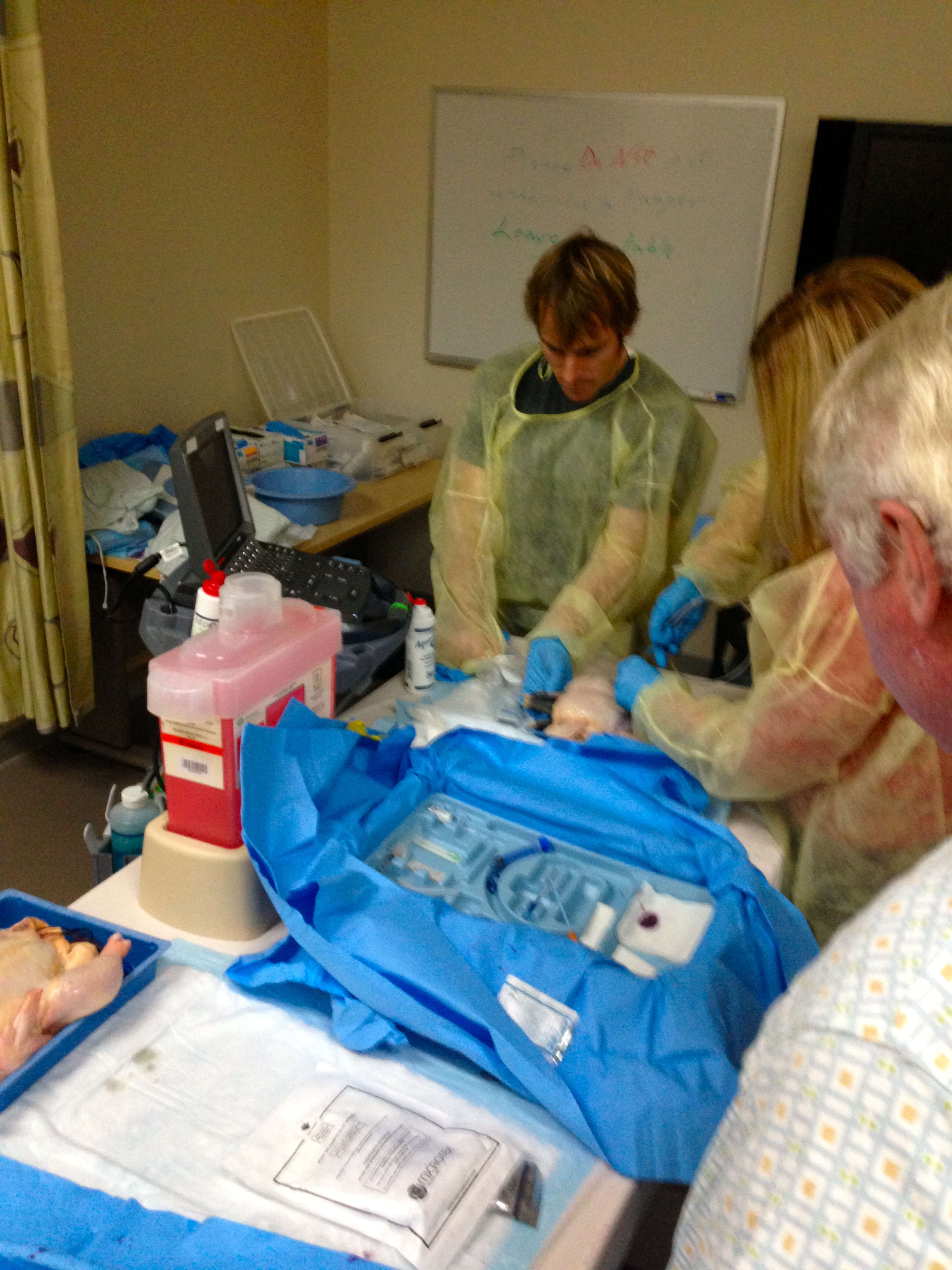
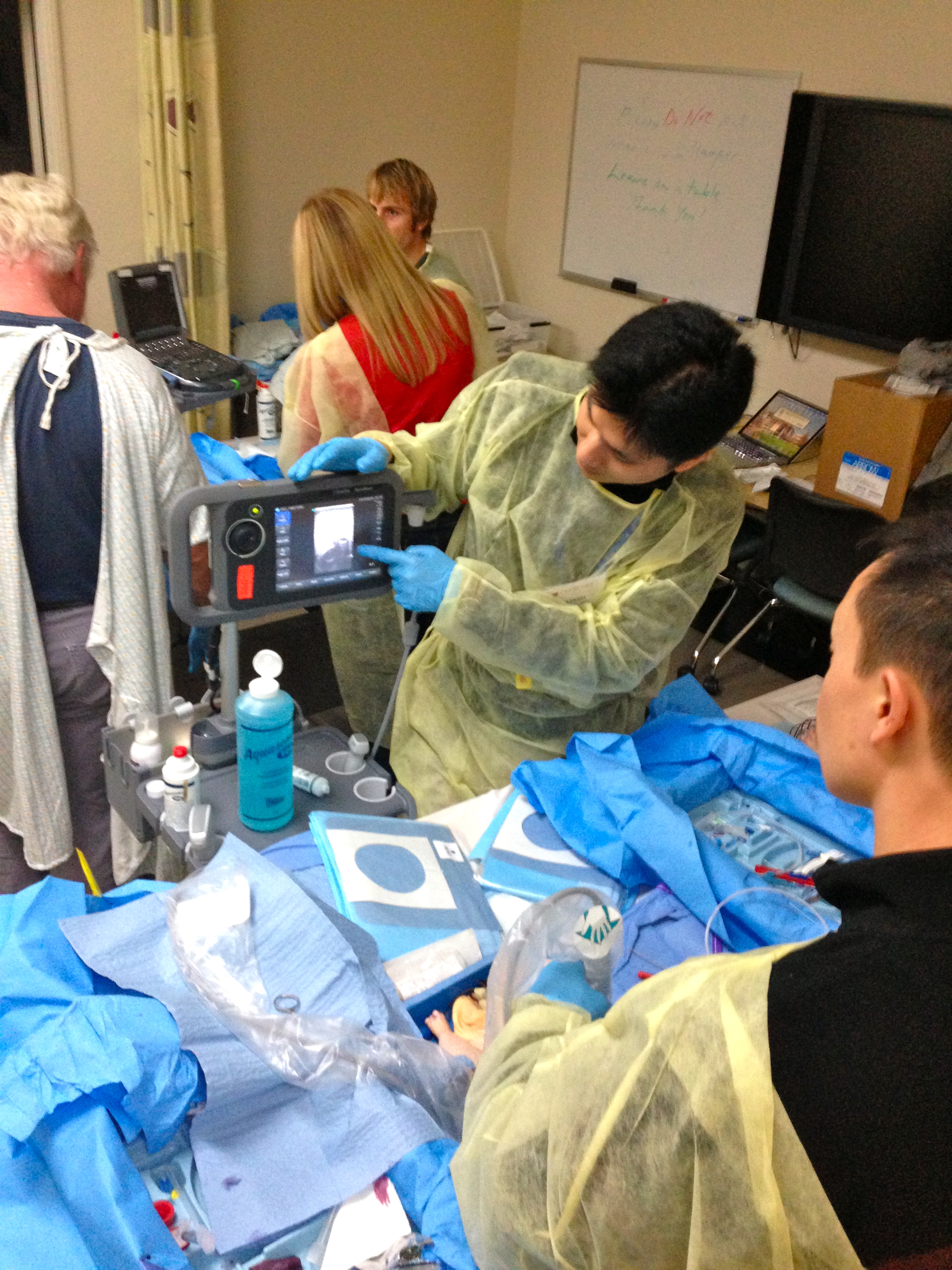
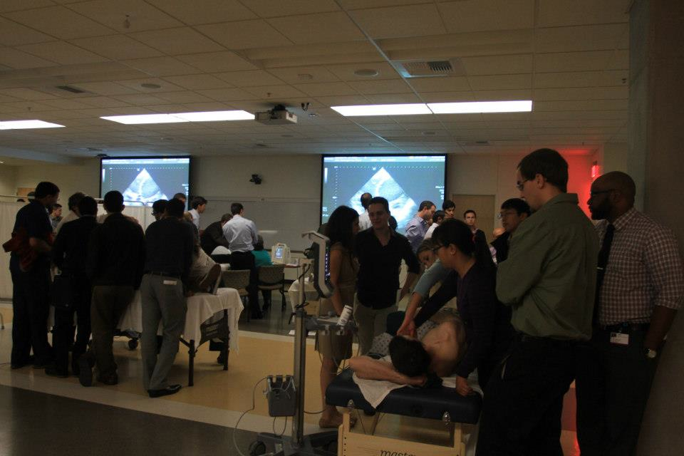
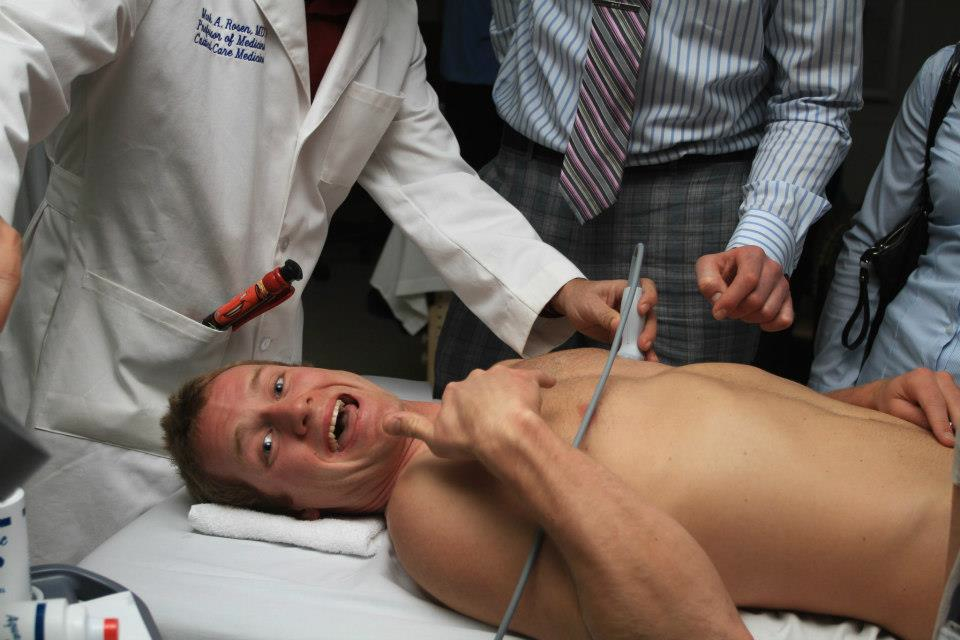
We then had 8 different hands-on stations with 11 different instructors (and 4 chickens used for central lines deserving of props, and lots of other simulators as you will see in the below pics)!  Including those described above, we also had the above instructors (from left to right, top to bottom (hyperlinks take you to “other” images that come up when you google search their names)- Dr. Viveta Lobo – our current US fellow and future Director of the Visiting Scholars Program, Dr. Brita Zaia – our past US fellow and current Kaiser San Francisco Ultasound Director, Dr. Darrel Sutijono – US fellowship trained faculty at Kaiser Santa Clara and new to twitter and #FOAMed and the FOAM movement, Dr. Manish Asarvala – US fellowship trained at UCSF and faculty at Kaiser Santa Clara. Dr. Yoshi Mitarai – an emergency medicine/critical care specialist who recently saved a life while at the gym doing Zumba (yup, you read that right), Dr. Suzanne Lippert – a specialist in nerve blocks and international/global health who is faculty at Stanford EM. Dr. Jennifer Newberry – an MD JD (so, super smart) and one of our senior EM residents who is staying on as a fellow in healthcare/ public policy.

IMG_1094IMG_1095 IMG_1097 IMG_1099 IMG_1100 IMG_1102 IMG_1103 IMG_1105

SonoMedStudent and UltraFest: Ultrasound in the Medical Schools…it’s time.

I just got back from the 2nd Annual UCI UltraFest – a FREE (yup, you read that right, FREE!) medical student ultrasound workshop held at UC Irvine this year for any and all medical students in California who want to come and learn bedside ultrasound using simulation and live models along with hearing about the future and international ultrasound. It’s getting a lot of press – as it should. It started off with an idea that came by one of my friends and mentors, Dr. Chris Fox (an ultrasound guru in his own right), and with the help of his medical students (Lancelot Beier, Kiah Bertoglio, and MaryJane Vennat), they organized faculty from all over California (UCI, Stanford, UCLA, USC) from multiple specialties. As Chris states “If I can give medical students the confidence and curiosity to want to use ultrasound all the time, then I feel they will take better care of their patients, and will provide more accurate care without turning to radiation.” You know how many medical students showed up? about 300. On a Saturday. During their free time. Some even wore a tie! …even when they ate from the free taco truck at lunch (yum!).

IMG_1084IMG_1077UltrafestpicUltrafestpic2ultrafestpic3For even more pics go here.

You know what that means? They want to learn it …and it should be incorporated into all medical school curriculums. It has been well studied to improve their learning in anatomy, physiology, and pathology… not to mention their patient care in their clinical years. It’s time. I’m excited to host it at Stanford for 2014!

The Dean of the School of Medicine at UC Irvine gave a quick speech welcoming the medical students about “Healthcare”: the practice, the word, and the meaning – and how that relates to ultrasound:

In a recent addition of the AAMC reporter, they discuss the FIRST ultrasound workshop held for the AAMC members and how medical students, like Kiah Bertaglio,  at UC Irvine, feel the need for ultrasound in medical education is a must. I posted about this fun AAMC event, that I was lucky enough to be a part of with some of my heroes, previously for your reading pleasure while SUSME and AIUM announced 2013 as the Year of Ultrasound (YOU) – highlighted by AIUM Ultrasound First group, the Life in the Fast Lane bloggers, the Ultrasound Podcast folks, and, of course, little ole’ me on SonoSpot while highlighting the ACEP US Section and the immense amount of social media interest/bloggers/tweets on the topic of bedside ultrasound.

In the AAMC Reporter. they state: “With rapid advancements in ultrasound technology, …a handful of the nation’s medical schools make ultrasound training a standard part of the curriculum. And there is a push to encourage more schools to use ultrasound….South Carolina is one of the first schools to implement a four-year interdisciplinary ultrasound curriculum. The program started in 2006 and is based on a training model for emergency medical workers. First- and second-year students learn how to read scans during lectures and lab sessions and through Web-based learning modules. In the third and fourth years, students use hand-held ultrasound devices to examine their first patients…..Richard Hoppmann, M.D., dean at South Carolina who also helped form SUSME, considers hand-held ultrasound devices the “stethoscopes of the 21st century.” “The technology is already here. What is lagging behind is the health care workforce who is knowledgeable and skilled in the appropriate use of these devices,” said Hoppmann, who stressed the importance of proper training….This portability allows doctors to perform bedside exams to detect acute emergencies such as internal bleeding, collapsed lungs, and intestinal obstructions. Ultrasound can be used to guide catheters with more accuracy, decreasing patient discomfort and saving time for staff. In addition, ultrasound is safer than other types of imaging because it does not emit potentially harmful radiation.

[A medical student said “It makes it exponentially easier to see real things, happening to real patients in real time. You are better able to identify something if you have seen it before.”] He added that seeing things in real time has a powerful effect on patients. Clarkson recalled one patient who, after viewing fluid buildup impairing his heart and kidneys on an ultrasound, realized he needed to improve his diet and take his medication….Like South Carolina, the University of California, Irvine, School of Medicine (UC Irvine), offers a four-year fully integrated ultrasound curriculum—the only one of its kind in California. What started in 2003 as a fourth-year elective in emergency ultrasound has expanded across the entire continuum….Students at UC Irvine have shown an extracurricular interest, organizing an Ultrasound in Medical Education Interest Group with guest lecturers and hands-on sessions. …Kiah Bertaglio, a third-year medical student at UC Irvine, helped arrange [UltraFest]. “The response was overwhelming and shows how important tomorrow’s doctors and health care workers see portable ultrasound becoming. It provided an incredible opportunity for students to learn and improve bedside ultrasound skills in multiple fields,” she said. Efforts to reach this goal are picking up at medical schools and teaching hospitals. The emergency medicine department at the Ohio State University Wexner Medical Center, East Virginia Medical School, and Wayne State University School of Medicine are incorporating ultrasound residency programs and fellowships. If this trend continues, Hoppmann predicts the technology could become a core competency that will enhance patient care across the board.”

SonoHangout & Journal Club: SonoSpot, BedsideSono, UltrasoundPodcast – the SMACC-down!

Get ready for the SMACC-down by some bedside ultrasound enthusiasts! What is SMACC, you ask? Well, if you don’t know, then make sure you visit THIS website. It’s the first conference of its kind, and even includes SonoWars as a very important part to help beat your fellow physician in various games of …well…..intellect (or, just allowing another reason why you can use the term SMACC-down – oh yeah!).
Now, we did our part in joining the SMACC club (yup, that’s right, an exclusive secret society whose membership provide secrets that you WISH you knew – ok, not really – but to find out more go here.) In this SMACC club entry, Drs. Mike Stone (@BedsideSono), Mike Mallin and Matt Dawson (ultrasound podcast guys), and little ole’ me discuss three hot (and I mean, HOT!!) articles, with visual effects that are sure to add us into the Oscar running for 2014 – this is sure to impress the judges! (they are in Australia….and New Zealand, in case you were wondering).
Download the articles prior to watching so you can follow along.  They are:

And, to get your SMACC on, see our SMACC down here:

SonoStudy and Review: The Beta hCG, the Ectopic, and the Ultrasound Findings – do they correlate?

In a recent article in the Journal of Ultrasound in Medicine (through AIUM), a study was done that illustrates exactly what we all experience in practice – an ectopic pregnancy can occur at any beta hCG level….AND a normal pregnancy can result despite a higher bHCG and no IUP seen. The conversations with the radiologists who still believe in “screening” who should and should not be scanned based solely on the beta hCG level will minimize – so we hope. The lowest beta hcg I have ever seen with a diagnosed ectopic? Brace yourselves…….152 ! There have been other case reports and cohort multi-site studies that you can read herehere, here, and here about low hCG and diagnosis of ectopic pregnancy. ACEP even has an article on it. But what if the beta hCG is high? …and you see nothing in the uterus on your ultrasound? There was a study done in 2011 by Wang, et al out of UCSF that discussed this too, asking if we should increase the discriminatory zone. There are also studies that show if you DO see something in the uterus, what does that mean in relation to ectopic pregnancy? Well, first, let’s talk physiology – Now, hCG is made by the syncytiotrophoblasts of the placenta after fertilization occurs, and correlates with the size and developing of the fetus…. well, Im going to stop there, as the only reason I stated that was to type “syncytiotrophoblasts” as I rarely have the opportunity to do so (insert sarcasm).

There is, however, a term used to describe the maternal serum hCG level above which a gestational sac should be consistently visible on transvaginal sonography – “discriminatory zone” – coined in the 1980s (yup, that’s right, 30 years ago!). This was thought to be 1,000, 1,500, or 2,000 on transvaginal ultrasound (and 3,600 or 6,000 on transabdominal ultrasound) depending on the study you read. So, if the hCG is above that zone and no IUP is seen – then you have yourself an ectopic pregnancy ….until proven otherwise! – and doctors would think treating for ectopic is the appropriate next step. Then there was a hiccup – There was a study that showed an HCG of 2,000 may not mean ectopic as 33% of the study’s subjects had a normal IUP after having no IUP on ultrasound when they were above that discriminatory zone. Oopsy! But, the prior studies all kinda had a possible gestational sac, but defined an IUP as the presence of a double decidua sign or yolks sac. So, this study wanted to know if there was no gestational sac and the bHCG was above this discriminatory zone, will there be an IUP, and if so, then what is the prognosis – in other words, is this discriminatory zone be valid?

“Objectives—The human chorionic gonadotropin (hCG) discriminatory level—the maternal serum β-hCG level above which a gestational sac should be consistently visible on sonography in a normal pregnancy—has been reported to be 1000 to 2000 mIU/mL for transvaginal sonography. We assessed whether a woman with a β-hCG above 2000 mIU/mL and no intrauterine fluid collection on transvaginal sonography can subsequently be found to have a live intrauterine gestation and, if so, what the prognosis is for the pregnancy.

 Methods—We identified all women scanned between January 1, 2000, and December 31, 2010, who met the following criteria: serum β-hCG testing and transvaginal sonography were performed on the same day; β-hCG was positive and sonography showed no intrauterine fluid collection; and a live intrauterine pregnancy was subsequently documented. We tabulated the β-hCG levels in these cases and assessed pregnancy outcome.

Results—A total of 202 patients met the inclusion criteria, including 162 (80.2%) who had β-hCG levels below 1000 mIU/mL on the day of the initial scan showing no intrauterine fluid collection, 19 (9.4%) with levels of 1000 to 1499, 12 (5.9%) 1500 to 1999, and 9 (4.5%) above 2000 mIU/mL. There was no significant relationship between initial β-hCG level and either first-trimester outcome or final pregnancy outcome (P> .05, logistic regression analysis and Fisher exact test). The highest β-hCG was 6567 mIU/mL, and the highest value that preceded a liveborn term baby was 4336 mIU/mL.

[Also: “Comparing outcomes in cases with β-hCG below 1000 versus above 1000 mIU/mL also showed no significant difference: 89.9% (125 of 139) live at the end of the first trimester in the low hCG group versus 88.6% (31 of 35) live in the high hCG group; 86.6% (110 of 127) liveborn in the low hCG group versus 80.6% (25 of 31) liveborn in the high hCG group (P > .05 for both comparisons Fisher exact test)]”

Conclusions—The hCG discriminatory level should not be used to determine the management of a hemodynamically stable patient with suspected ectopic pregnancy, if sonography demonstrates no findings of intrauterine or ectopic pregnancy.

New Guidelines published in NEJM in Oct 2013 have changed the criteria in order to reduce the risk of prematurely stating a pregnancy is non-viable.

 A great discussion on this also heard here BroomeDocs’ Casey Parker: here

For a great 5 minute talk on Ectopic pregnancy and how to identify it by ultrasound, see Dr. Phil Perera’s Soundbytes insert – but, as the studies above suggest, if you see no IUP despite an hCG above the discriminatory zone, there may not be an ectopic pregnancy – make sure to look around the adnexal region, and have close follow up with the Ob/Gyn doctor.

SonoStudy & Review of literature: Rapid Lung/cardiac/IVC – differentiates causes of acute dyspnea

A recent study in cardiovascular medicine … a concept that has been highlighted in varying ways from prior studies (by Liteplo (ETUDES study), Lichtenstein (all of his studies, actually), Volpicelli (ILC-LUS international consensus), and Manson with the RADIUS study/protocol), continues to conclude that rapid bedside ultrasound of lung/cardiac/IVC can help differentiate causes of acute dyspnea. The state: “The present study demonstrated that rapid evaluation by lung-cardiac-inferior vena cava (LCI) integrated ultrasound has a higher diagnostic accuracy for differentiating acute dyspnea due to AHFS from pulmonary acute dyspnea (including COPD/asthma, pulmonary fibrosis, and ARDS) compared with lung ultrasound either alone or in combination with plasma BNP assay. These findings suggest that LCI integrated ultrasound has become a fundamental tool for diagnostic evaluation of patients with acute dyspnea and selection of early treatment in the emergency setting.”

The algorithm below is what they used:

1476-7120-10-49-1

ABSTRACT:

BACKGROUND: Rapid and accurate diagnosis and management can be lifesaving for patients with acute dyspnea. However, making a differential diagnosis and selecting early treatment for patients with acute dyspnea in the emergency setting is a clinical challenge that requires complex decision-making in order to achieve hemodynamic balance, improve functional capacity, and decrease mortality. In the present study, we examined the screening potential of rapid evaluation by lung-cardiac-inferior vena cava (LCI) integrated ultrasound for differentiating acute heart failure syndromes (AHFS) from primary pulmonary disease in patients with acute dyspnea in the emergency setting.

METHODS:

Between March 2011 and March 2012, 90 consecutive patients (45 women, 78.1 +/- 9.9 years) admitted to the emergency room of our hospital for acute dyspnea were enrolled. Within 30 minutes of admission, all patients underwent conventional physical examination, rapid ultrasound (lung-cardiac-inferior vena cava [LCI] integrated ultrasound) examination with a hand-held device, routine laboratory tests, measurement of brain natriuretic peptide, and chest X-ray in the emergency room.

RESULTS:

The final diagnosis was acute dyspnea due to AHFS in 53 patients, acute dyspnea due to pulmonary disease despite a history of heart failure in 18 patients, and acute dyspnea due to pulmonary disease in 19 patients. Lung ultrasound alone showed a sensitivity, specificity, negative predictive value, and positive predictive value of 96.2, 54.0, 90.9, and 75.0%, respectively, for differentiating AHFS from pulmonary disease. On the other hand, LCI integrated ultrasound had a sensitivity, specificity, negative predictive value, and positive predictive value of 94.3, 91.9, 91.9, and 94.3%, respectively.

CONCLUSIONS:

Our study demonstrated that rapid evaluation by LCI integrated ultrasound is extremely accurate for differentiating acute dyspnea due to AHFS from that caused by primary pulmonary disease in the emergency setting.

SonoCase: 35 yr old – acute urinary retention, penile pain – by Drs. Peabody, Mailhot, Perera

In the recent issue of WestJEM, a case report of another excellent application for bedside ultrasound is described by our very own Dr. Phil Perera (yup, he is more than just the RUSH exam). A video where he discusses the ultrasound application and case follows….

“A 35-year-old man presented to the emergency department (ED) for acute urinary retention and penile pain for 4 hours. The patient denied any significant medical history or history of trauma. Physical exam revealed testicles that were nontender, without masses. However, a tender mass was felt at the distal end of the penis, adjacent to the urethral meatus. Placement of a Foley catheter resulted in a return of 700 cc of clear yellow urine and immediate resolution of the patient’s suprapubic and penile pain.

During the ED course, the Foley catheter was removed with a subsequent trial of voiding. Initially, the patient was able to void 15 cc of urine until the normal stream was abruptly cut off. The patient then complained of extreme penile pain, near the urethral meatus. A small, circular and firm mass was again palpated in the distal penile shaft. Bedside emergency ultrasound (EUS), performed with a 10 MHz linear array probe placed along long axis of penis, revealed a hyperechoic, dense and round structure with characteristic acoustic shadowing at the distal end of the urethra, with obstruction of the urinary flow (Video). The object, a 9 mm stone, was removed with forceps. Following stone removal, the patient experienced immediate pain relief and was able to spontaneously void.

While urethral imaging has traditionally been performed with retrograde urethrography (RUG), more recently ultrasound has been used to minimize the pain associated with RUG and to provide clinicians more detailed information about urethral pathology.2 As demonstrated in this case, EUS allowed a prompt diagnosis of the patient’s condition with appropriate rapid treatment and removal of the urethral stone.”

SonoStudy: >12,000 kids – Identifying factors putting kids at low risk, not needing CT after trauma

There has been quite a bit of press lately on this –  Here and Here – And for good reason. With the ALARA principle, and being a pediatric population which has been studied so many times with regard to trauma and the need for CT, a recent study by Holmes et al published in Annals of Emergency Medicine did a multi-site study enrolling >12,000 kids and identified 7 factors that places children at very low risk for injury not requiring abdominal CT. A prior post discusses a study done by the same author and my thoughts of pediatric US in trauma. BTW – Dr. Holmes also discusses low risk factors for adult patients in a prior study too.

The prediction rule for pediatric patients consisted of (in descending order of importance):
No evidence of abdominal wall trauma or seat belt sign,
Glasgow Coma Scale score greater than 13,
No abdominal tenderness,
No evidence of thoracic wall trauma,
No complaints of abdominal pain,
No decreased breath sounds, and
No vomiting.

Now, I dont know about you, but to me it is quite obvious – we just now have a nicely powered study that we can use for all the doctors who want to CT despite all of the above being negative. The authors say that if any one of the above exist then a decision by the physician should be made as to what the next best management step would be – observation period with serial exams, ultrasound (holla!), CT – are all options depending on clinical judgement. Below is the abstract:

Study objective: We derive a prediction rule to identify children at very low risk for intra-abdominal injuries undergoing acute intervention and for whom computed tomography (CT) could be obviated.
Methods: We prospectively enrolled children with blunt torso trauma in 20 emergency departments. We used binary recursive partitioning to create a prediction rule to identify children at very low risk of intra-abdominal injuries undergoing acute intervention (therapeutic laparotomy, angiographic embolization, blood transfusion for abdominal hemorrhage, or intravenous fluid for ≥2 nights for pancreatic/gastrointestinal injuries). We considered only historical and physical examination variables with acceptable interrater reliability.
Results: We enrolled 12,044 children with a median age of 11.1 years (interquartile range 5.8, 15.1 years). Of the 761 (6.3%) children with intra-abdominal injuries, 203 (26.7%) received acute interventions. The prediction rule consisted of (in descending order of importance) no evidence of abdominal wall trauma or seat belt sign, Glasgow Coma Scale score greater than 13, no abdominal tenderness, no evidence of thoracic wall trauma, no complaints of abdominal pain, no decreased breath sounds, and no vomiting. The rule had a negative predictive value of 5,028 of 5,034 (99.9%; 95% confidence interval [CI] 99.7% to 100%), sensitivity of 197 of 203 (97%; 95% CI 94% to 99%), specificity of 5,028 of 11,841 (42.5%; 95% CI 41.6% to 43.4%), and negative likelihood ratio of 0.07 (95% CI 0.03 to 0.15).
Conclusion: A prediction rule consisting of 7 patient history and physical examination findings, and without laboratory or ultrasonographic information, identifies children with blunt torso trauma who are at very low risk for intra-abdominal injury undergoing acute intervention. These findings require external validation before implementation.

SonoStudy: Emergency Physicians can estimate gestational age in 1st trimester pregnancies

I recently posted about how emergency physicians can decrease the length of stay of patients with first trimester vaginal bleeding/pelvic pain (ruling out ectopic pregnancy) by performing a bedside pelvic ultrasound, which also had a couple cases to ponder about. As emergency physicians are getting more and more savvy with bedside ultrasound, it may benefit the patient’s future care if we are also able to tell them the gestational age. Well, this study (and great review) on emergency physicians-performed ultrasound estimating gestational age (compared with radiology results) highlights exactly that! And, guess what? we CAN estimate gestational age – shocking, I know. Below is the abstract:

ABSTRACT:

BACKGROUND: Patient reported menstrual history, physician clinical evaluation, and ultrasonography are used to determine gestational age in the pregnant female. Previous studies have shown that pregnancy dating by last menstrual period (LMP) and physical examination findings can be inaccurate. Radiology department ultrasound has proven to be the most accurate way of determining gestational age. The aim of this study is to determine the accuracy of emergency department ultrasound as an estimation of gestational age (EDUGA) in an emergency department (ED) population.

METHODS:

A prospective convenience sample of ED patients presenting in the first trimester of pregnancy (based upon self-reported LMP) regardless of their presenting complaint were enrolled. EDUGA was compared to gestational age estimated by ultrasound performed in the department of radiology (RGA) as the gold standard. Pearson’s product moment correlation coefficient was used to determine the correlation between EDUGA compared to RGA.

RESULTS:

Sixty-eight pregnant patients presumed to be in the 1st trimester of pregnancy based upon self-reported LMP consented to enrollment. When excluding the cases with no fetal pole, the median discrepancy of EDUGA versus RGA was 2 days (interquartile range (IQR) 1 to 3.25). The correlation coefficient of EDUGA with RGA was 0.978. When including the six cases without a fetal pole in the data analysis, the median discrepancy of EDUGA compared with RGA was 3 days (IQR 1 to 4). The correlation coefficient of EDUGA with RGA was 0.945.

CONCLUSION:

Based on our comparison of EDUGA to RGA in patients presenting to the ED in the first trimester of pregnancy, we conclude that emergency physicians are capable of accurately performing this measurement. Emergency physicians should consider using ultrasound to estimate gestational age as it may be useful for the future care of that pregnant patient.

SonoStudy: Flat IVC predictor of poor prognosis in trauma – A-B-C-D-Echo in Trauma!

A recent study on the IVC and trauma and acute surgical patients was done by Ferrada et al, and despite the giggles I get when I read it and how they describe the IVC as “Fat ” or “Flat”, it is an important topic to discuss as it is one of the few articles out there that correlate the iVC to trauma patients and acute surgical patients. First off, it is a retrospective study, which can make it difficult to assess patients with similar factors without other factors coming into play (but when is that NOT the case, honestly?) They did compare the IVC in all patients and studied those patients who seemed sick as well – ICU admission, immediate surgery need, transfusion needed. The power of the study was good but not great – 101 patients studied – varying in type of trauma and surgical need. There was a previous study published in the Journal of Trauma in 2011 that stated CT evidence of flat IVC was an indicator for hypovolemia and  poor prognostic indicator for blunt solid organ injuries – this confirmed a study done in 2010 stating the same thing. Thankfully, ultrasound can get you that information much more immediately than CT!

This month, another study by the same author (Ferrada) in the Journal of trauma and acute care surgery entitled A-B-C-D-Echo (I know, love it!) stated that adding limited transthoracic echo, including the IVC, will benefit trauma patients with results showing “Flat inferior vena cava was associated with an increased incidence of ICU admission (p < 0.0076) and therapeutic operation (p < 0.0001). Of the 148 patients, 27 (18%) had LTTE results indicating euvolemia. The diagnosis in these cases was head injury (n = 14), heart dysfunction (n = 5), spinal shock (n = 4), pulmonary embolism (n = 3), and stroke (n = 1). Of the patients, 121 had LTTE results indicating hypovolemia. Twenty-eight hypovolemic patients had a negative or inconclusive Focused Assessment with Sonography for Trauma examination finding (n = 18 penetrating, n = 10 blunt), with 60% having blood in the abdomen confirmed by surgical exploration or computed tomographic scan. Therapy was modified as a result of LTTE in 41% of cases. Strikingly, in patients older than 65 years, LTTE changed therapy in 96% of cases.”

Below are the Abstracts of the studies highlighted by Ferrada:

“Flat inferior vena cava (IVC) on ultrasound examination has been shown to correlate with hypovolemic status. We hypothesize that a flat IVC on limited echocardiogram (LTTE) performed in the emergency room (ER) correlates with poor prognosis in acutely ill surgical patients. We conducted a retrospective review of all patients undergoing LTTE in the ER from September 2010 until June 2011. IVC diameter was estimated by subxiphoid window. Flat IVC was defined as diameter less than 2 cm. Fat IVC was defined as diameter greater than 2 cm. Need for intensive care unit admission, blood transfusion requirement, mortality, and need for emergent operation between patients with flat versus Fat IVC were compared. One hundred one hypotensive patients had LTTE performed in the ER. Average age was 38 years. Admission diagnosis was blunt trauma (n = 80), penetrating trauma (n = 13), acute care surgery pathology (n = 7), and burn (n = 1). Seventy-four patients had flat IVC on initial LTTE. Compared with those with fat IVC, flat patients were found have higher rates of intensive care unit admission (51.3 vs 14.8%; P = 0.001), blood transfusion requirement (12.2 vs 3.7%), and mortality (13.5 vs 3.7%). This population also underwent emergent surgery on hospital Day 1 more often (16.2 vs 0%; P = 0.033). Initial flat IVC on LTTE is an indicator of hypovolemia and a predictor of poor outcome.”

ABCDEcho:

“BACKGROUND: Limited transthoracic echocardiogram (LTTE) has been introduced as a technique to direct resuscitation in intensive care unit (ICU) patients. Our hypothesis is that LTTE can provide meaningful information to guide therapy for hypotension in the trauma bay.

METHODS: LTTE was performed on hypotensive patients in the trauma bay. Views obtained included parasternal long and short, apical, and subxyphoid. Results were reported regarding contractility (good vs. poor), fluid status (flat inferior vena cava [hypovolemia] vs. fat inferior vena cava [euvolemia]), and pericardial effusion (present vs. absent). Need for surgery, ICU admission, Focused Assessment with Sonography for Trauma examination results, and change in therapy as a consequence of LTTE findings were examined. Data were collected prospectively to evaluate the utility of this test.

RESULTS: A total of 148 LTTEs were performed in consecutive patients from January to December 2011. Mean age was 46 years. Admission diagnosis was 80% blunt trauma, 16% penetrating trauma, and 4% burn. Subxyphoid window was obtained in all patients. Parasternal and apical windows were obtained in 96.5% and 11%, respectively. Flat inferior vena cava was associated with an increased incidence of ICU admission (p < 0.0076) and therapeutic operation (p < 0.0001). Of the 148 patients, 27 (18%) had LTTE results indicating euvolemia. The diagnosis in these cases was head injury (n = 14), heart dysfunction (n = 5), spinal shock (n = 4), pulmonary embolism (n = 3), and stroke (n = 1). Of the patients, 121 had LTTE results indicating hypovolemia. Twenty-eight hypovolemic patients had a negative or inconclusive Focused Assessment with Sonography for Trauma examination finding (n = 18 penetrating, n = 10 blunt), with 60% having blood in the abdomen confirmed by surgical exploration or computed tomographic scan. Therapy was modified as a result of LTTE in 41% of cases. Strikingly, in patients older than 65 years, LTTE changed therapy in 96% of cases.

CONCLUSION: LTTE is a useful tool to guide therapy in hypotensive patients in the trauma bay.”