In the most recent issue of EP Monthly, Drs. Teresa Wu and Brady Pregerson, give another enlightening and humorous story of the average every-day emergency patient after first describing how they explained to their EM intern the role of emergency physicians (which I couldnt agree more with!): “As an EM physician, you are a healer, an educator, a detective, a diagnostician, and a master strategist all rolled into one.”
The case:
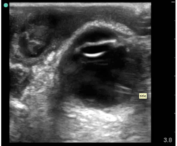
“41-year-old male who presents to the ED with concerns that his left eye is progressively getting more swollen. He’s had some increasing eye pain and purulent drainage over the past six days. At first he thought that he was just having really bad seasonal allergies, but today, he started feeling a “pulling sensation” on the medial aspect of his left eye. He denies any headache, diplopia, sinus pain, rhinorrhea, nausea, vomiting, or recent trauma. He does note a subjective fever at home, and his temperature is 38.2°C in the ED. His vital signs otherwise demonstrate tachycardia to 123 bpm, but a normal blood pressure, oxygen saturation, and respiratory rate. Your intern has asked the nurse to obtain a visual acuity on the patient and he is systematically going through his ocular exam when you walk by the room to check on him. He comes out of the room to give you an update on what he’s discovered so far. The patient has tenderness to palpation over his left medial orbit and possible entrapment on ocular exam. He has no additional pain with extraocular movement and no diplopia, but has so much periorbital edema that it wasn’t possible to get a consistent Tonopen measurement. There doesn’t appear to be any fluorescein uptake on the slit lamp exam, and other than conjunctival injection and the lid swelling, the patient has a normal ocular exam.
It is now about 4:30 pm and you know that in 30 minutes, all consultants turn into pumpkins and their pagers magically stop working. As you are about to ask your intern what he wants to do next, the medical student pulls up the ultrasound machine that the intern asked her to wheel over and hands it to him. He takes the linear array transducer and performs an ocular ultrasound at the bedside. He saves the following images: what do you see?”
To find out…..and read up on what it was, what happened, and the pearls of the exam, go here.
Merry Christmas everyone! For your reading pleasure this week, Id thought we would discuss a case whose topic is near and dear to my heart. In the most recent issue of EPMonthly, there is a great case and interesting “internal” discussion made quite humorously public by Drs. Pregerson and T. Wu of a young healthy male with right lower quadrant abdominal pain after eating at a “Roach Coach”…. which just so happen to have the best breakfast burritos, but I digress… The case discussion involves how the history and physical may help, how labs may (or may not) help and how an ultrasound can be of use to make you and your surgical colleagues feel better in taking the patient to the OR. There was a recent post on SonoSpot about ultrasound in appendicitis sharing data from a study about the CT findings when US “equivocal” cases arise. When the ultrasound is positive – how great is that?! Quite a few studies recently on the topic and some of the more recent ones can be found here.
The case is followed by an extensive (and great) discussion of the technique, pearls and pitfalls of ultrasound in evaluating the appendix – because we all know there are quite a few. As far as the sensitivity ad specificity go, they state it best:
“Sensitivity & Specificity: Both the sensitivity & specificity of ultrasound for appendicitis are less than that of CT. In pediatrics the values are about 88% and 94% respectively, and in adults about 83% and 93%. (These numbers may vary depending on the experience of the ultrasonographer.) There are studies from Europe and Israel where they have used the “ultrasound first” approach for many, many years that show even better test characteristics. These values are actually not that bad when compared to CT scan whose sensitivity and specificity are around 94% and 95% respectively. Remember, however, that the performance characteristics for ultrasound can be significantly worse in overweight patients or those with overlying bowel gas. In addition, if the appendix is retrocecal or is lying in a difficult anatomical plane, the study will be more challenging. Unfortunately, you may still have to do a CT scan if your ultrasound is non-diagnostic and your clinical suspicion is moderate to high, but the strategy of ultrasound first would likely decrease CTs by about 50%.”
And in kids…”You should be aware of the most recent recommendation of the American College of Radiology from the “Choosing Wisely” campaign, which states, “Don’t do computed tomography (CT) for the evaluation of suspected appendicitis in children until after ultrasound has been considered as an option.” Although CT is accurate in the evaluation of suspected appendicitis in the pediatric population, ultrasound is nearly as good in experienced hands. Since ultrasound will reduce radiation exposure, ultrasound is the preferred initial consideration for imaging examination in children. If the results of the ultrasound exam are equivocal, it may be followed by CT. This approach is cost-effective, reduces potential radiation risks and has excellent accuracy, with reported sensitivity and specificity of 94 percent.”
To diagnose appendicitis: look for a noncompressible a-peristaltic structure that attaches to the cecum that is larger than 7mm in diameter.
A great tutorial of ultrasound for the appendix can be found here by the UltrasoundPodcast guys:
I’d like to introduce everyone to our amazing Ultrasound Fellow, Dr. Viveta Lobo (otherwise known as “VLo” to our team – of course!). She came to us by way of Drexel, tolerates out antics, appreciates our quirks, and laughs at our jokes. We love her! Enjoy this post about a healthy guy who looked very sick, short of breath, and only bedside ultrasound, using the RADIUS protocol, could help diagnose it so quickly and get the patient what he needs and fast….
I’m about 4 months into my Ultrasound (US) Fellowship at Stanford, and while I am thrilled to have greatly improved my US skills, and image acquisition during a scanning shift, it is in no comparison to the thrill, and satisfaction I felt, after using my bedside US skills to navigate through the following case.
A 55-year-old healthy male, with no past medical history, presents with progressively worse shortness of breath over the past 2 weeks. Within 30 seconds of being in the room, he is getting more short of breath, dusky, diaphoretic, and requiring to now sit up and lean forward while speaking to me in 1 word sentences. He is on a 100% non re-breather, sating about 93%. The rest of his vitals – BP 124/84 RR 41 HR 124 Temp 97.8
Even as a new attending, I was pretty certain, that if I did not figure this out in the next few minutes, this once very healthy patient is going to decompensate, and likely end up with grave morbidity. However, given that he had no known history, I had nothing to go by, except…. I grabbed my US probe, and within 3 minutes, I gained a wealth of information. I first took a look at his chest by using the phased array low frequency probe on each side of his chest in 8 total areas (4 on each side). This is what I see throughout:
…. >2 large B lines bilaterally, rays from the pleural line on the top to the end of the screen.
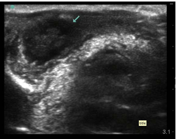
When I switched to a RUQ and LUQ views, my suspicions were confirmed :
RUQ:
LUQ:
…Now, the US images are on cardiac presets so the resolution is a touch different than what we are used to, but the findings are obvious which heightened my concern for the patient even more: large pleural effusions noted bilaterally. Seen as a black (anechoic) area above the diaphragm. Black is fluid on ultrasound, and you can even see the lung trying to breathe on each of the images above.
Next, I quickly assessed his IVC, and saw a plump dilated IVC, consisted with fluid overload state, which prompted me to stop my nurse from hanging any IV fluids. I then performed a bedside echo:
Subxiphoid view: (placing the phased array low frequency probe in the subxiphoid area and pressing down while flattening the probe, using the liver as an acoustic window to see the heart):
Apical 4-chamber view: (placing the probe just underneath the nipple line, at the point of maximal impulse and angling toward the body center):
…. I was able to rule out a pericardial effusion and cardiac tamponade as well as any significant RV strain to suggest a hemodynamically unstable pulmonary embolism, but I appreciated significant left ventricle dysfunction, and hypokinesis.
I then took a look using the linear probe on the anterior chest wall at the 2nd intercostal space and saw:
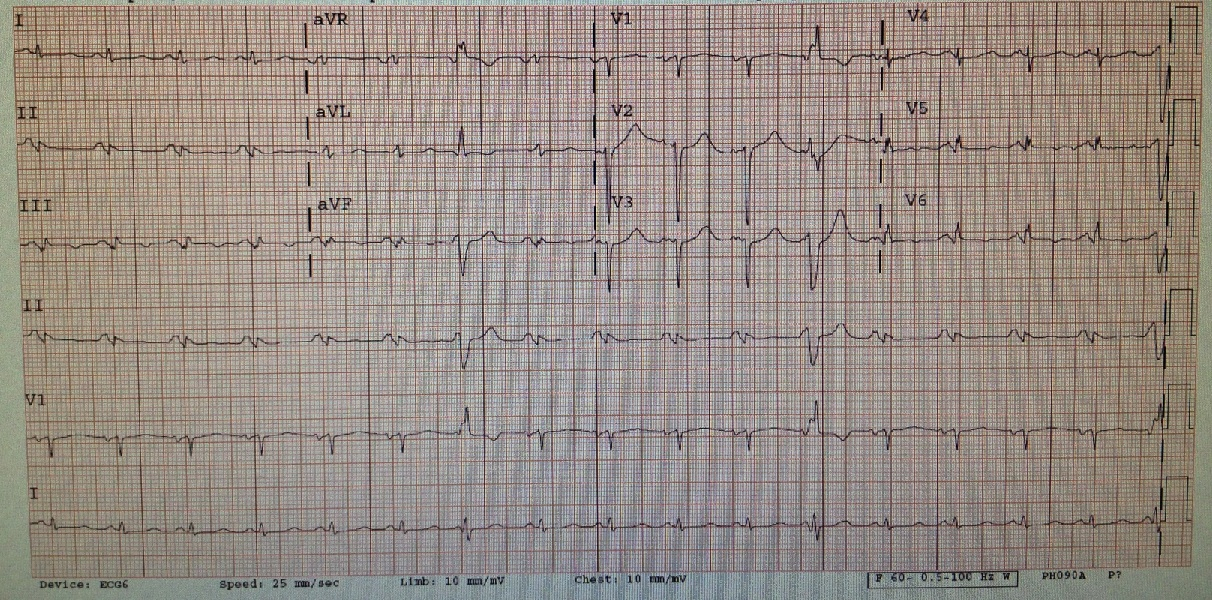
So, there is great lung sliding but we see it almost too well! The reason is because fluid is the lover of ultrasound and will allow you to see tissue deep to it better due to enhancing of echoes. There is fluid between the parietal and pleural layers, more and more from superior to inferior chest – on both sides. That’s quite a bit of pleural effusion if it goes all the way up to the upper lung zones! While I was putting the pieces together and realizing the diagnosis, my nurse informs me that his istat troponin comes back elevated. His initial EKG:
…..showed sinus tachycardia with ischemic changes inferior and laterally, with t waves inversions. We also see multiple PVCs. No old one EKG for comparison. Ah, the evolution of an MI on EKG – love it!
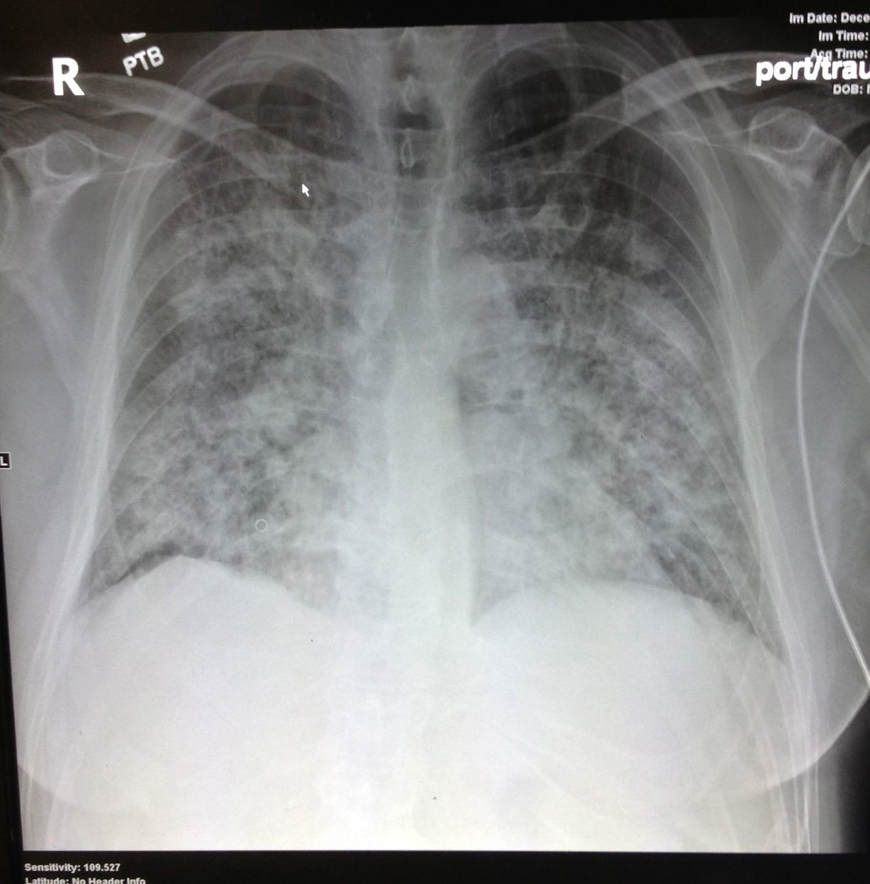
So to recap, I have an otherwise healthy gentlemen, with progressive sob, no chest pain, but with positive family history of ACS, with confirmed LV dysfunction on US and bilateral pleural effusions and a positive troponin, and some possible ischemic changes on EKG. Sounds like a post ischemic cardiac event presenting with ventricular infarct! From door to diagnosis in 5 minutes! I placed him on BIPAP, gave him a big shot of Lasix IV, aspirin PO, and called my cardiologist! The patient started to improve after the medication, avoiding intubation. The Chest Xray was then done:
….showing bilateral diffuse opacities which could be typical for ARDS.
After a brief cardiology evaluation, my patient was admitted to the CCU and shortly after went to the Cath lab, and was found to have a complete LAD occlusion.
While I initially had a very broad differential including PE, new onset CHF, cardiac tamponade, myocarditis, pneumonia; my bedside ultrasound was quickly able to prioritize my differential, and consult the right service, with a specific question of – should this patient go to the cath lab? Without bedside US, this patient could have easily been a Medical ICU evaluation for respiratory distress, with an extensive work up, including CT Chest, intubation, and more time than the patient needed for a diagnosis to have bee made while we sorted through the differential.
This case is one of many that completely validates bedside ultrasound for me, and my decision to pursue this awesome fellowship!
As a follow up: Patient went on to get an LVAD, and is on the heart transplant list.
In the most recent issue of EPMonthly, our good friend, Dr. Teresa Wu, and Brady Pregerson right up a case they had of a patient with abdominal pain. In their wisest and most sarcastic way, they present this case with a great teaching point (ok, there are many teaching points as you will find on the last page of the case – but one in particular that deserves special mention). Read on and see if you can get what that point may be…
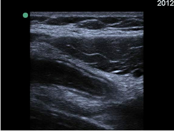
“56-year-old otherwise healthy female who presented to the ED with a chief complaint of “severe abdominal pain” after she finished lifting boxes of heavy books at her job the day before. She states her pain is worse with movement and is better when she lies still. She has never had pain like this before, and today, it is 10 out of 10 in severity. The pain is described as sharp and tearing, but it does not radiate to her chest or back. She has no other associated symptoms, and she has tried Ibuprofen without any relief.
Her vital signs are all completely within normal limits and her physical exam is only remarkable for tenderness to palpation over her left rectus muscles, and a seemingly pulsatile aorta palpable through her thin abdominal wall. She has no rebound or guarding on abdominal exam, and she has no other abnormal findings. Given her symptoms and her palpable aorta, your senior resident decides it would be prudent to do a quick scan of her aorta to make sure nothing catastrophic is imminent.” The following image was obtained:”
The Aorta seems ok. Hmmmm…..Still wonder what happened to the case and what it was? Read the issue in depth and you’ll then get to know and love Teresa Wu as much as I do.
Hint – look at the entire screen when evaluating any organ by bedside ultrasound…..
In the most recent installment of the Sound Judgement Series by AIUM, Drs. Rochelle F. Andreotti and Sara M. Harvey from the Department of Radiology at Vanderbilt discuss the use, accuracy and effectiveness of ultrasound for acute pelvic pain. It seems that pelvic pain has, again, become an important issue as there are quite a few articles that have come out about it recently, likely because there are so many visits to clinics and emergency departments with this exact chief complaint. As the authors state “The diagnosis can be challenging because many symptoms and signs lack sensitivity and specificity. Urgent life-threatening conditions requiring surgical intervention (eg, ectopic pregnancy, appendicitis, a ruptured ovarian cyst, and ovarian torsion) and fertility-threatening conditions (eg, pelvic inflammatory disease [PID] and ovarian torsion) should take precedence over other disorders.” – Guess which imaging modality can evaluate all of them? Continue reading →
An article that just recently came to my attention made me start to think a little bit about how we teach how to do the FAST scan. In a prior post, I discuss the RUQ and LUQ details – to ensure to not miss any amount of free fluid that should be seen on the FAST scan, keeping in mind it’s limitations. Then, I read this article in the EMJ online First from April 2012 that discusses a case of an ‘unusually’ positive FAST scan, but when reading about the injury and the location, I am not surprised about the location of free fluid development. Hind-sight is 20/20, but it highlights a few key concepts that should always be addressed: look for free fluid in the REGION on the RUQ and LUQ, not only between the liver/spleen and kidneys AND serial FAST scans for any patient where the mechanism suggests a risk for intra-abdominal injury (particularly if you are not going to CT the patient) – I do this frequently in the patients who come in drunk as all get-out where I cannot rely on my physical exam or the pediatric population where radiation would be best avoided if possible.
This case is one of those cases that make me so proud of the residents I work with…. Drs. Brianne Steele and Cesar Avila identified the need for a RUSH exam, but didn’t stop there – they noticed something during their RUSH and proceeded with another evaluation – obtaining the surprising diagnosis below, saving him time in the emergency department and canceling his CT scan that didn’t need to be done, which I then conclude controls his healthcare charge. period.
This is a guest post from my good friend and colleague, Dr. Zoe Howard, an ultrasound lover and user, part of ACEP’s medical student initiative, and helping us incorporate bedside ultrasound into the medical school curriculum. She had an amazing case where bedside ultrasound helped make the correct diagnosis for a patient who was getting worse, bounced back to the ED, and stayed in an observation unit to be seen by her (and the ultrasound machine) in the morning:
A sweet 15yo girl presented with a week of suprapubic pain and dysuria… Continue reading →
The great thing about bedside ultrasound is that you can get a really REALLY good idea of what is going on with a patient within 5-10 minutes of their arrival, particularly patients who can’t tell you whats going on (whether it’s because they are lethargic and tachypneic – like this case – or altered, unconscious, or speak another language) , but, because you are a great doc, you do know by just walking through the doorway and looking at the patient that he is S.I.C.K. This case discusses exactly that and highlights the RUSH protocol, (see my prior post on the evidence based approach to the RUSH) ,but also how interpreting those applications when correlating to your exam and clinical history is key and adds greatly to your evaluation of the patient.
60 yr old guy (with an amazingly nice wife and family) with a history of cutaneous T-cell lymphoma (chemo/radiation 3 months earlier), Sezary syndrome (with chemo) and Sjogren’s syndrome walks in (yes, thats right, walks in…) to the emergency department waiting room, leaning on his wife after just getting off a plane from Seattle (about a 3 hour flight) after a 1 week cruise. Continue reading →
This case was quite interesting and a great pick-up by the EM resident, Dr Cesar Avila. It highlights the use of ocular ultrasound with eye complaints/vision change/trauma, especially when you cannot properly evaluate the eye well due to eyelid swelling.
82 year old male with history of globe rupture and retinal detachment status post repair two months earlier presents to the ED with eyelid swelling of that same eye, gradual onset over one day with now inability to open eyelids well with yellow discharge coming from eye. Continue reading →